2678
Tailored SEMs for wave modulations in SMS imaging1Department of Radiology, Medical Physics, Medical Center, University of Freiburg, Faculty of Medicine, Freiburg, Germany
Synopsis
The use of a matrix gradient coil enables to tailor spatial encoding magnetic Fields (SEMs) for slice specific frequency shifts. Applying such shifts in oscillatory manner allows for novel methods of signal separation in SMS imaging.
Introduction
Multiple parallel imaging techniques based on the spatial varying receive RF-senstivity profiles have been presented to speed-up the inherently slow MR-image acquisition[1,2]. Additional methods to increase the separability between multiple slices are usually based on slice-dependent phase shifts[3-5]. Deploying a matrix gradient coil [6-9] enables to tailor specific spatial encoding magnetic fields (SEMs) for a slice-dependent frequency manipulation, as presented at last years ISMRM [10]. A complete signal separation by frequency shifts becomes unattainable for high resolutions, which requires very strong non-linear SEMs. Here we present a method which deploys oscillating frequency shifts which are constant within each slice and alternate in time which relates to an apparent oscillatory movement along the frequency encoding direction. Utilizing the same frequency with a phase shift or a different oscillatory-frequency in other slices enhances the signal separation between multiple slices.Methods
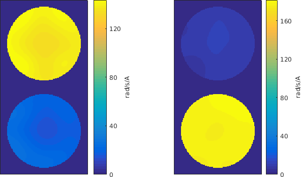
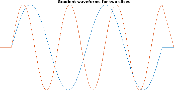
Applying a constant SEM during read out enlarges the $B_0$-field and shifts all signals within this slice along the read-out direction. Opposing constant fields in different slice positions shift signals in opposite directions and may be used to enhance signal separability for SMS-imaging [10]. A full signal separation is possible, however limited by the available strength and dephasing effects which arise from the field imperfections. Oscillation of a constant SEM during readout spreads the signal in k-space similar to Fronsac [11] or wave-caipi [5]. Oscillations which are either phase-shifted or with different oscillation frequencies allow for even better signal separation in SMS-imaging. Additionally, application of constant SEMs with same amplitude variations as for phase-encoding induces an additional slice dependent phase-shift. Additional SEMs are required to be constant throughout one slice and zero throughout others. A Tikhonov-regularized L2 based optimization was used to find suitable current distributions and clustered matrix coil channels. Measured SEMs used to induce frequency shifts in two slices are depicted in Fig.1 along with a corresponding exemplary trajectory. Experiments were performed on a 3T scanner (Siemens Trio, a Tim system) equipped with additional gradient insert hardware which was developed and built in-house consisting of the following components: 84-channel matrix gradient coil [2-4]; controller, clock distribution board, 12-channel digital-to-analog converter [6] and a 12-channel gradient power amplifier setup (IECO, XPA-150-350). The gradient coil was clustered to 12 channels, each containing four or eight elements from one ring. Pulse sequences were programmed in Pulseq [7] to simultaneously control the MR scanner and the additional hardware. Simulations, pulse sequence programming and data processing was performed in Matlab (Natick, USA). Demonstration experiments were acquired using a gradient echo sequence with TE/TR of 15/200ms and a flip angle of 20°.Results
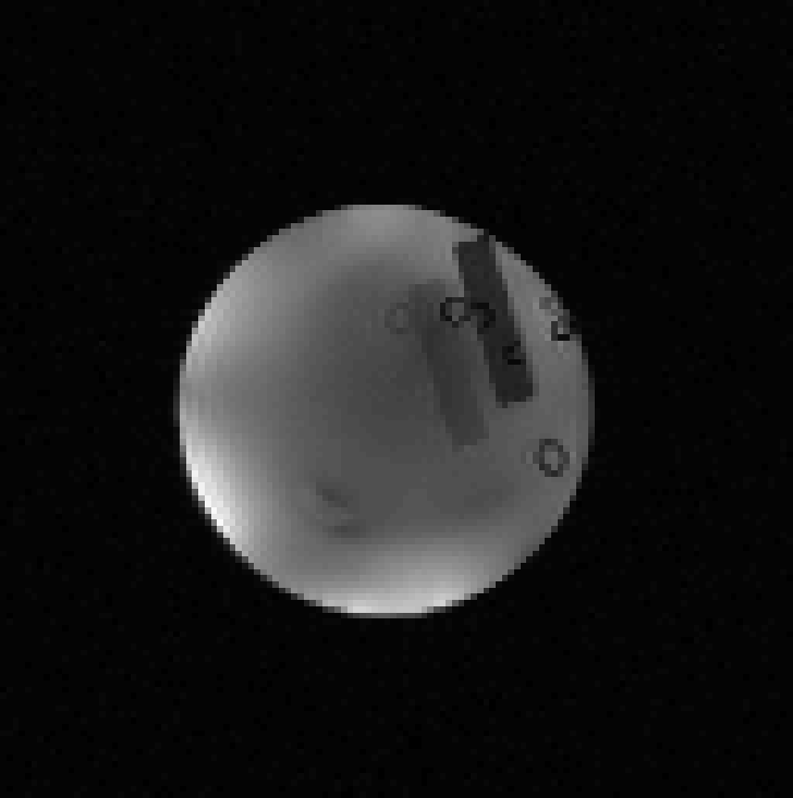
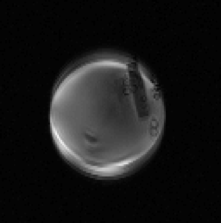
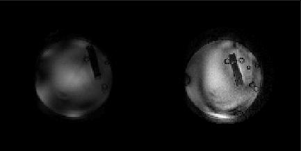
A basic image without aliasing of two overlaying slices is shown in Fig.3. Aliasing from applying the additional SEMs during read-out is depicted in Fig.4. The standard k-space formalism is not sufficient to describe the resulting aliasing. It may rather be compared to a fast movement of the object which is equivalent to spreading signals over more then one k-space point. By using high oversampling this additional information can be extracted. Reconstructed images of the two slices are shown in Fig.5. All features can be uncovered. However, some residual distortions are not removed.Discussion
New degrees of freedom offered by a matrix gradient coil may be used for novel ways of signal manipulation in SMS-imaging. The ability to tailor SEMs in a slice dependent manner offers new degrees of freedom beyond the traditional k-space formalism. However, due to comparably high frequency manipulations an accurate timing of the hardware is crucial. Further increase in image quality is expected from the use of magnetic field monitoring technology [12,13] to achieve accurate time and magnitude calibration of the modulating fields.Acknowledgements
This work was supported by the European Research Council grant 282345 ’RANGEmri’.References
[1] Griswold, M. A., Jakob, P. M., Heidemann, R. M., Nittka, M., Jellus, V., Wang, J., Kiefer, B. and Haase, A. (2002), Generalized autocalibrating partially parallel acquisitions (GRAPPA). Magn. Reson. Med., 47: 1202–1210. doi:10.1002/mrm.10171
[2] Pruessmann, K. P., Weiger, M., Scheidegger, M. B. and Boesiger, P.
(1999), SENSE: Sensitivity encoding for fast MRI. Magn. Reson. Med., 42:
952–962.
[3] Breuer, F. A., Blaimer, M., Heidemann, R. M., Mueller, M. F., Griswold, M. A. and Jakob, P. M. (2005), Controlled aliasing in parallel imaging results in higher acceleration (CAIPIRINHA) for multi-slice imaging. Magn. Reson. Med., 53: 684–691. doi:10.1002/mrm.20401
[4] Setsompop, K., Gagoski, B. A., Polimeni, J. R., Witzel, T., Wedeen, V. J. and Wald, L. L. (2012), Blipped-controlled aliasing in parallel imaging for simultaneous multislice echo planar imaging with reduced g-factor penalty. Magn. Reson. Med., 67: 1210–1224. doi:10.1002/mrm.23097
[5] Bilgic, B., Gagoski, B. A., Cauley, S. F., Fan, A. P., Polimeni, J. R., Grant, P. E., Wald, L. L. and Setsompop, K. (2015), Wave-CAIPI for highly accelerated 3D imaging. Magn. Reson. Med., 73: 2152–2162. doi:10.1002/mrm.25347
[6] Littin, et al. (2015), Shielded Matrix Gradient Coil. Proc. 23th ISMRM Congress, Toronto (2015): 1022.
[7] Littin, et al. (2016), Implementation of an 84 Channel Actively Shielded Matrix Gradient Coil. Proc. 24th ISMRM Congress, Singapore (2016): 3561.
[8] Littin, S., Jia, F., Layton, K. J., Kroboth, S., Yu, H., Hennig, J. and Zaitsev, M. (2017), Development and implementation of an 84-channel matrix gradient coil. Magn. Reson. Med.. doi:10.1002/mrm.26700
[9] Jia, F., Littin, S., Layton, K. J., Kroboth, S., Yu, H., Hennig, J. and Zaitsev, M. (2017), Design of a shielded coil element of a matrix gradient coil, Journal of Magnetic Resonance, Volume 281, 2017, Pages 217-228, ISSN 1090-7807, https://doi.org/10.1016/j.jmr.2017.06.006.
[10] Littin, et. al., (2017), Simultaneous Multi Slice Imaging Using Matrix Gradient Coils. Proc. 25th ISMRM Congress, Hawaii (2017): 0517.
[11] Wang, H., Tam, L. K., Constable, R. T. and Galiana, G. (2016), Fast rotary nonlinear spatial acquisition (FRONSAC) imaging. Magn. Reson. Med., 75: 1154–1165. doi:10.1002/mrm.25703
[12] Barmet, C., Zanche, N. D. and Pruessmann, K. P. (2008), Spatiotemporal magnetic field monitoring for MR. Magn. Reson. Med., 60: 187–197. doi:10.1002/mrm.21603
[13] Testud, F., Gallichan, D., Layton, K. J., Barmet, C., Welz, A. M.,
Dewdney, A., Cocosco, C. A., Pruessmann, K. P., Hennig, J. and Zaitsev,
M. (2015), Single-shot imaging with higher-dimensional encoding using
magnetic field monitoring and concomitant field correction. Magn. Reson.
Med., 73: 1340–1357. doi:10.1002/mrm.25235
Figures