2673
T1-weighted bipolar fat/water separated spin-echo PROPELLER acquired with dual bandwidths1Department of Clinical Neuroscience, Karolinska Institutet, Stockholm, Sweden, 2Neuroradiology, Karolinska University Hospital, Stockholm, Sweden, 3GE Healthcare, Stockholm, Sweden
Synopsis
A bipolar fat/water separated T1-weighted dual-bandwidth spin-echo PROPELLER sequence is proposed which achieves strong and homogenous fat suppression without any dead time. Dual bandwidth sequences are compared against a corresponding fat saturated sequence in terms of SNR and CNR efficiency.
Purpose
The PROPELLER technique offers a motion robust alternative to the Cartesian spin echo sequence and has been extended to enable separation of fat and water (1–4). Since every PROPELLER blade samples the k-space center, several low-resolution images of the prescribed field of view are acquired, which enables blade-wise fat/water separation (3).
For two-point spin echo acquisitions, using readouts with different bandwidths (5), the dead time associated with shifted readouts is removed, increasing the SNR efficiency (4). To encode chemical shift, the second readout bandwidth is either increased or decreased to acquire the signal before or after the second spin echo (4).
We propose a T1-weighted fat/water separated PROPELLER sequence with dual bandwidths, where bipolar gradients are used instead of a second refocusing pulse. This enhances the contrast between gray and white matter otherwise degraded by magnetization transfer effects. We compare the two dual bandwidth sequences in terms of SNR and CNR efficiency.
Methods
A T1-weighted PROPELLER sequence (ETL = 1) with dual bandwidths (4) was implemented according to Figure 1. In the bipolar mode, the second refocusing pulse was removed and the second readout changed polarity. The encoding patterns for the dual-bandwidth acquisitions are displayed in Figure 2.
Fat/water separation was performed bladewise in k-space (6), with field map estimation using graph cuts (7) and initial phase estimation (8). After field map and initial phase demodulation, water and fat were estimated using the regularized pseudoinverse of the model matrix $$$\mathbf{A}$$$ (6, 9), defined as:
$$\mathbf{A}^{\dagger}_k = (\mathbf{A}_k^H \mathbf{A}_k+\kappa_k \mathbf{I})^{-1}\mathbf{A}_k^H,$$
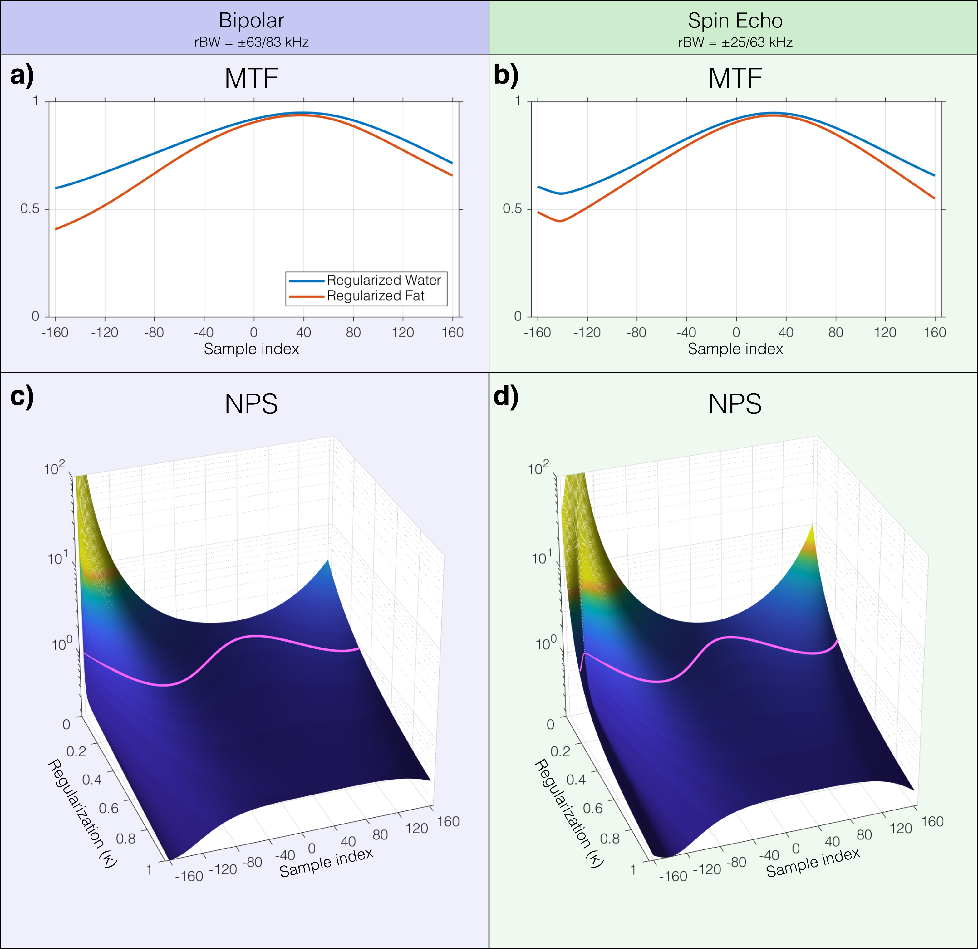
where the Tikhonov regularization factor $$$\kappa$$$ was varied between k-space points k (9). We propose to adapt $$$\kappa$$$ to achieve a uniform noise power spectrum (NPS). The modulation transfer function (MTF) and noise amplification factor for water and fat was calculated from the diagonal elements of $$$\mathbf{A}_k^\dagger\mathbf{A}_k$$$ and $$$tr(\mathbf{A}_k^\dagger (\mathbf{A}_k^\dagger)^H)$$$, respectively.
To allow for SNR and CNR measurements, the sequence was further modified so that each phase encoding line was acquired twice before acquiring the next line. ROI’s were placed in gray matter, white matter, and cerebrospinal fluid. SNR efficiency is the SNR divided by the square root of the sequence duration (including fat saturation if applicable).
Experiments were performed on a volunteer on a 1.5T clinical MRI system (DVMR450, GE Healthcare) using an eight-channel head coil. Data was acquired with the following scan parameters: TR = 500 ms, minimum TE, 16 blades, matrix size = 320×32, 11 slices with 3 mm thickness, R=1, FOV = 240mm, TA = 4min 15s (×2 for SNR measurements). Echo shift Δ and rBW are given in figures. Fat saturation PROPELLER data with similar scan parameters and acquisition time was also acquired for comparison.
Results
MTF and NPS for different amount of Tikhonov regularization are shown in Figure 3. The NPS is equalized by applying the contoured kappa (magenta line in Figure 3c-d).
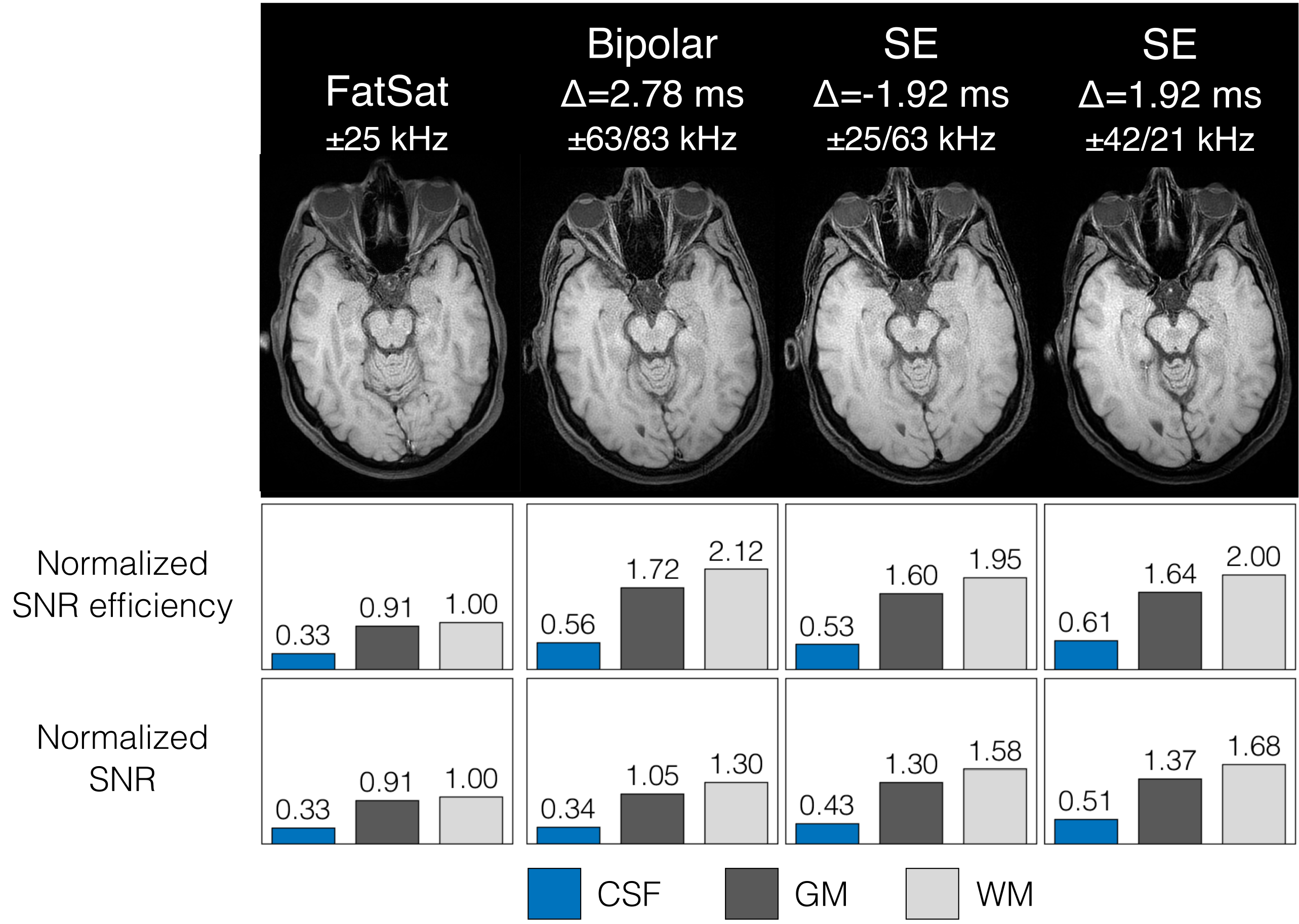
Regularized water (NPS=1) images of the optical nerve with corresponding SNR and SNR efficiency measurements are shown in Figure 4, normalized to WM in the fat saturation acquisition.
Discussion & Conclusion
In addition to providing images with and without fat suppression, the proposed bipolar and spin echo sequences are superior to fat saturation in several aspects; the optical nerve is clearly visible due to the strong fat suppression, and both SNR and GM/WM CNR is higher. The increased efficiency is expected as the duration of fat saturation itself penalizes the SNR efficiency. The SNR is higher in the spin echo images compared to bipolar, which is expected since the bandwidths are lower. However, the efficiency is slightly higher in the bipolar case, indicating that it is a strong option in situations where many slices are required and TR is too short for low bandwidth acquisitions. The higher CNR efficiency in the bipolar image is likely explained by the removal of the second refocusing pulse and its associated magnetization transfer effects. The image quality in Figure 4 is degraded by motion due to the long acquisition time necessary for SNR evaluation.
Long readout durations are associated with poor conditionality of the signal model at certain points in k-space. Even though the readout gradient durations are shorter with the present bipolar protocol, regularization was required to reduce colored noise, as is seen in Figure 3. The cost of equalizing the NPS is a non-uniform modulation transfer function (MTF), shown in Figure 3a-b. Given well designed scan parameters, the suppressed spatial frequencies will be in the outer parts of k-space where the energy typically is lower.
In conclusion, we have presented a bipolar dual bandwidth fat/water separated spin echo PROPELLER sequence that offers strong and homogenous fat suppression with high SNR and CNR efficiency.
Acknowledgements
No acknowledgement found.References
- Huo D, Li Z, Aboussouan E, Karis JP, Pipe JG. Turboprop IDEAL: A motion-resistant fat–water separation technique. Magn. Reson. Med. 2009;61:188–195.
- Weng D, Pan Y, Zhong X, Zhuo Y. Water–fat separation with parallel imaging based on BLADE. Magn. Reson. Imaging 2013/6;31:656–663.
- Schär M, Eggers H, Zwart NR, Chang Y, Bakhru A, Pipe JG. Dixon water-fat separation in PROPELLER MRI acquired with two interleaved echoes. Magn. Reson. Med. 2016;75:718–728.
- Rydén H, Berglund J, Avventi E, Norbeck O, Skare S. T1-weighted two-point fat/water separated PROPELLER acquired with dual bandwidths. In: Proceedings of the 25th Annual Meeting of ISMRM, Hawaii, USA. ; 2017. p. 1017.
- Eggers H. Variable Bandwidth Turbo Spin-Echo Dixon Imaging. In: Proceedings of the 23rd Annual Meeting of ISMRM, Milan, Italy. ; p. 1657.
- Brodsky EK, Holmes JH, Yu H, Reeder SB. Generalized k-space decomposition with chemical shift correction for non-Cartesian water-fat imaging. Magn. Reson. Med. 2008;59:1151–1164.
- Berglund J, Skorpil M. Multi-scale graph-cut algorithm for efficient water-fat separation. Magn. Reson. Med. [Internet] 2016. doi: 10.1002/mrm.26479.
- Bydder M, Yokoo T, Yu H, Carl M, Reeder SB, Sirlin CB. Constraining the initial phase in water-fat separation. Magn. Reson. Imaging 2011;29:216–221.
- Lu W, Yu H, Shimakawa A, Alley M, Reeder SB, Hargreaves BA. Water-fat separation with bipolar multiecho sequences. Magn. Reson. Med. 2008;60:198–209.
Figures
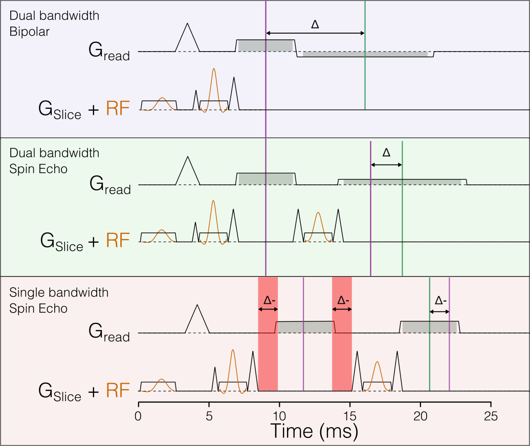
Figure 1 - Pulse sequence diagrams of the investigated methods. Magenta lines mark spin echo and green lines mark the center of the readout. The dual bandwidth design removes the dead time of shifted design (red patches).