2666
Distribution-controlled and optimally spread non-Cartesian sampling curves for accelerated in vivo brain imaging at 7 Tesla1CEA/NeuroSpin - INRIA/Parietal, Gif-sur-Yvette, France, 2CNRS - ITAV, Toulouse, France, 3Siemens Healthineers, Saint-Denis, France, 4CEA/NeuroSpin/UNIRS/METRIC, Gif-sur-Yvette, France
Synopsis
This work reports the use of new non-Cartesian k-space trajectories whose improved efficiency allows to significantly reduce MR scan time with minimum deterioration of image quality. Instead of using simple geometrical patterns, we introduce an approach inspired from stippling techniques, which automatically designs optimized sampling patterns along any distribution by taking full advantage of the hardware capabilities. Our strategy leads to drastically accelerated acquisitions, as demonstrated by our experimental results at 7T on in vivo human brains. We compare our method to widely-used non-Cartesian trajectories (spiral,radial) and demonstrate its superiority regarding image quality and robustness to system imperfections.
Introduction
Reducing scan times in Magnetic Resonance Imaging (MRI) is essential to explore higher spatial resolution which could help to better diagnose certain pathologies such as Alzheimer's disease. Methods to accelerate MRI such as parallel imaging [1] or compressed sensing [2] commonly rely on simple sampling patterns such as straight lines, spirals or slight variations of these elementary shapes. Such strategies do not take full advantage of the degrees of freedom offered by the hardware. In addition, they cannot be easily adapted to fit arbitrary sampling distribution and lack robustness at high acceleration factors (AF). Here, we report the use of a method called SPARKLING (Spreading Projection Algorithm for Rapid K-space sampLING), which may overcome these limitations by taking a completely different approach to the design of faster k-space sampling. A key ingredient underlying our approach is to take advantage of a shorter dwell time to acquire more points in a given observation window. In addition, this method relies on optimization to automatically generate non-Cartesian k-space trajectories under the hardware constraints, to fulfill 3 key criteria for optimal sampling: (i) a controlled distribution (e.g. along a variable density), (ii) a locally uniform coverage and (iii) a controlled distance between consecutive samples on a sampling curve. Preliminary results respecting the criteria (i)-(ii) were presented for ex vivo acquisitions in [3] but were unable to produce superior results as their sampling rate was too small.To illustrate SPARKLING advantages over standard non-Cartesian sampling, we used the proposed patterns to accelerate the fully-sampled Cartesian reference acquisition and compared it with equivalently accelerated variable-density spiral and radial acquisitions. These T2*-weighted 2D brain images were acquired on 4 healthy volunteers with a 7T scanner for high in plane resolution (390㎛x390㎛) and various acceleration factors up to 20.Materials and Methods
Setup
Four healthy volunteers were scanned with a 7T system (Siemens Healthineers,Erlangen,Germany) and a 1Tx/32Rx head coil (Nova Medical,Wilmington,MA,USA). Maximum gradient and slew rate magnitudes on this scanner were respectively 50 mT/m and 333 T/m/s and the gradient raster time was 10㎲. All subjects signed a written informed consent form and was enrolled in the study under the approval of our institutional review board.
Sequence and k-space trajectories
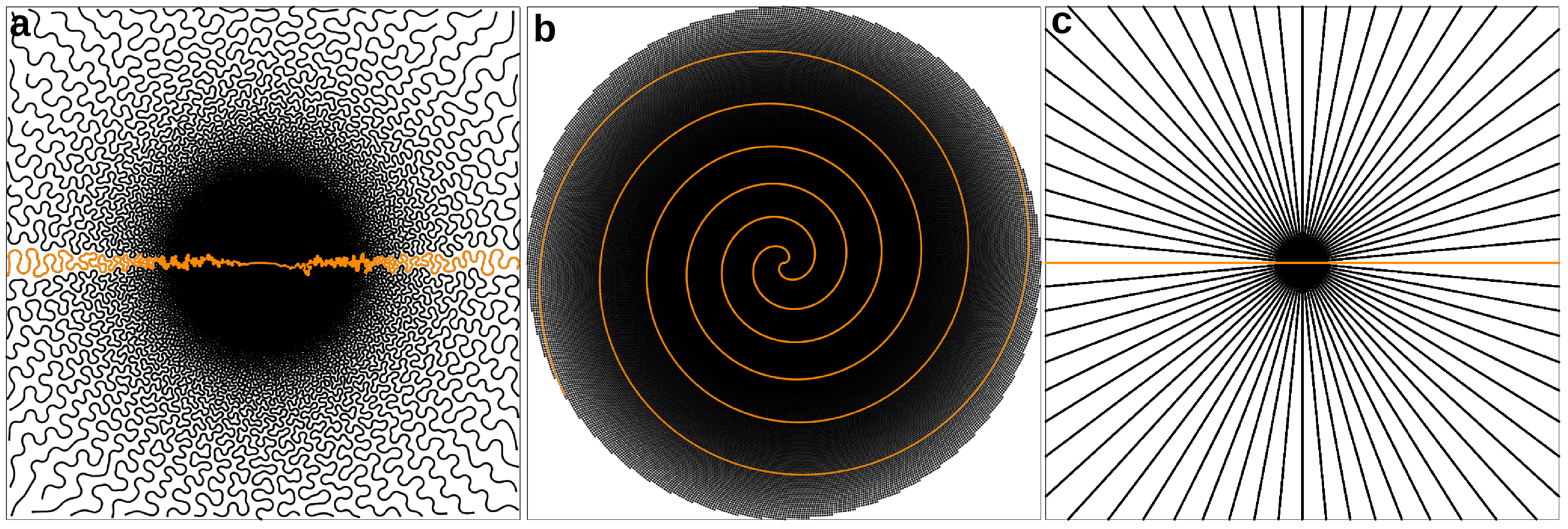
A modified 2D T2*-weighted GRE sequence was acquired for an in plane resolution of 390㎛ with the following parameters: TR=550 ms, TE=30 ms and FA=25° for one transversal slice of 3mm. Acquisitions were performed using the SPARKLING trajectories for different acceleration factors of 10, 15 and 20. (Fig.1a) displays a 15-fold accelerated SPARKLING trajectory segmented into 34 symmetric shots each acquiring 3072 samples during a readout time of 30.72 ms. The SPARKLING sampling was distributed along a radially decaying density. Limits in gradient and slew rate amplitudes were respectively set to 40 mT/m and 200 T/m/s. Total acquisition time (TA) was 28s, which is 15 times faster than the fully-sampled acquisition of 512 Cartesian lines (TA=4min42s). For comparison, we acquired a variable-density spiral (Fig.1b) [4] and a radial trajectory (Fig.1c) with the same numbers of shots, samples and TA as SPARKLING patterns.
Image reconstruction
Images were reconstructed using a self-calibrating nonlinear algorithm minimizing a sparsity promoting regularized CS-SENSE (Compressed Sensing SENsitivity Encoding) criterion in the wavelet domain similar to [5,6,7] which was adapted to non-Cartesian samples using the NFFT [8].
Results
The reconstructed brain images and a detail (yellow square) are presented in (Fig.2) for the fully sampled Cartesian reference acquisition (TA=4min42s) in (a-b). The second row displays the SPARKLING reconstructions for acceleration factors of AF=10 (c-d), AF=15 (e) and AF=20 (f). The third row displays the variable-density spiral reconstructions for AF=10 (g-h), AF=15 (i) and AF=20 (j). The fourth row displays the radial reconstructions for AF=10 (k-l), AF=15 (m) and AF=20 (n). The image quality is visibly stable across growing acceleration factors for the SPARKLING acquisitions and the latter manages to preserve fine structural details such as small vessels. On the contrary, important distortions due to off-resonance effects are visible on the spiral reconstructions while the undersampled radial patterns resulted in an overly smoothed image presenting streaking artifacts at AF=20.Discussion and conclusion
Here, we demonstrated the in vivo superiority of the proposed SPARKLING sampling over standard Cartesian, radial and spiral approaches. Although accelerated by a factor of 20, SPARKLING acquisitions were able to preserve fine structural details and proved to be more robust to system imperfections. Most interestingly, our method can adapt to any desired sampling density, observation window, MR weighting and hardware specifications. Although we focused on 2D acquisitions as a proof-of-concept, our technique can be extended to 3D acquisitions for which further gains are anticipated. Our findings may be used to accelerate numerous MR acquisitions from anatomical to dynamic imaging [9].Acknowledgements
We would like to thank Nicolas Boulant for his valuable advice. This research program was supported by DRF Impulsion grant in 2016 (COSMIC, P.I.: P.C.). C.L. was also supported by the CEA international PhD program.References
[1] : Pruessmann KP, Weiger M, Scheidegger MB, Boesiger P. (1999). SENSE: sensitivity encoding for fast MRI. Magnetic resonance in medicine. 42(5):952-62.
[2] : Lustig M, Donoho D, Pauly JM. (2007). Sparse MRI: The application of compressed sensing for rapid MR imaging. Magnetic resonance in medicine. 58(6):1182-95.
[3] : Lazarus C, Weiss P, Chauffert N, Mauconduit F, Bottlaender M, Vignaud A, Ciuciu P. (2017). SPARKLING: Novel non-Cartesian sampling schemes for accelerated 2D anatomical imaging at 7T using compressed sensing. 25th annual meeting of the International Society for Magnetic Resonance Imaging.
[4] : Lee JH, Hargreaves BA, Hu BS, Nishimura DG. (2003). Fast 3D imaging using variable‐density spiral trajectories with applications to limb perfusion. Magnetic resonance in medicine. 50(6):1276-85.
[5] : Wu B, Millane RP, Watts R, Bones P. (2008). Applying compressed sensing in parallel MRI. Proceedings of the 16th Annual Meeting of ISMRM (Vol. 1480).
[6] : Liu B, Zou YM, Ying L. (2008). SparseSENSE: application of compressed sensing in parallel MRI. Information Technology and Applications in Biomedicine. ITAB 2008. International Conference (pp. 127-130).
[7] : Boyer C, Ciuciu P, Weiss P, Mériaux S. (2012). HYR 2 PICS: Hybrid regularized reconstruction for combined parallel imaging and compressive sensing in MRI. Biomedical Imaging (ISBI), 2012 9th IEEE International Symposium (pp. 66-69).
[8] : Keiner, J., Kunis, S., and Potts, D. (2009). Using NFFT 3 - a software library for various nonequispaced fast Fourier transforms. ACM Trans. Math. Software, 36 : Article 19, 1-302.
[9] : Feng L, Axel L, Chandarana H, Block KT, Sodickson DK, Otazo R. (2016). XD‐GRASP: Golden‐angle radial MRI with reconstruction of extra motion‐state dimensions using compressed sensing. Magnetic resonance in medicine. 75(2):775-88.
Figures