2658
Blurring and Ghosting Effects Under Beats Formation in Magnetic Resonance Imaging Under Source Vibration1Electrical Engineering and Computer Science, Massachusetts Institute of Technology, Cambridge, MA, United States, 2Information Systems Technology and Design, Singapore University of Technology and Design, Singapore, Singapore, 3Engineering Product Development, Singapore University of Technology and Design, Singapore, Singapore
Synopsis
A key challenge of MRI is development of an accurate model of noise generation which are integral to generation of high-resolution images. Currently, motion induced noise is addressed at algorithmic level. Here, we present a novel physical model which incorporates the role of mechanical vibration of body parts in generation of additional frequency components in the emitted radio frequency spectrum around the precession frequency. The mathematical model was validated through a computational simulation which led to the discovery that beats generated as a result of mechanical vibrations of the source lead to ghosting and blurring effects.
Introduction
Enhanced signal to noise ratio leads to high resolution of images in MRI [1]. Noise in MRI has been studied from the perspective of thermodynamic fluctuations on magnetization and its relaxation [2] which tend to influence precession and relaxation [3] along with spatial drift of nuclei [4]. Motion of the subject is also an additional source of noise which is addressed at algorithmic level [5] . However, the fundamental aspect of additional frequency components generated as a consequence of mechanical vibration of the sample remains unexplored. The objective of current work is development and validation of a novel model of generation of noise in MRI under the mechanical vibration of the source,which results in additional spectrum components around the precession frequency which blurs the final image.
Methods
In an MRI system, the magnetic moment oscillates with a precession frequency, ω0=γB0, where γ is the gyromagnetic ratio and B0 is the applied magnetic flux density. The magnetic flux density B at a distance r from a source in a medium with permeability μ0 having a magnetic moment m oscillating at a frequency ω is expressed as. B(r)=(µ0)/(4π)(msinωt)/r3.. If the initial position of the magnetic moment shows a periodic spatial shift at a frequency ωm, at velocity defined by v=v=vmsinωt, the value of r can be found by integrating v with respect to time and it will have an impact on the generated magnetic field. The net change in magnetic flux density at a distance r can be expressed as follows,
B(r,t)=(µ0)/(4π)(vmsinωmt,)(3mcosω0t)/r4. It can be simplified as, B(r,t)=(3µ0)/(4πr4)mvm[cos(ωmt-ω0t)-cos(ωmt+ω0t)] which implies beats formation. The additional frequencies generated has corresponding impact on the magnetization and the relaxation times T1 and T2 in the Z and XY plane respectively.
For Hydrogen ions at 1 T, the precession frequency is 42.574MHz [1]. During imaging, if the source is vibrating at a frequency of 1kHz, which corresponds to a period of 1 ms , an additional frequency band would be observed in the output image. This source vibration are caused by movement of different body parts like vocal tract, respiratory system,or unintended body part movement by the subject.
Simulation and Results
We developed a mathematical model while incorporating the effect of mechanical vibrations of the source on the magnetic moment in MRI systems. The decay of magnetization in the XY plane under beats formation can be expressed as, MXY=MXY0e-t/T2 [cos(ωmt-ω0t)-cos(ωmt+ω0t)], where MXY0 is the initial value of magnetization in the XY plane. Similarly, the decay of magnetization in the Z plane is MZ=MZ0e-t/T1 [cos(ωmt-ω0t)-cos(ωmt+ω0t)], where MZ0 is the initial value of magnetization in the Z plane.
This model introduces the effect of source vibration on the MRI image reconstruction from the quantitative data (T1 and T2) collected from MRI machine of strength 1 Tesla and precession frequency of 42.576 MHz.
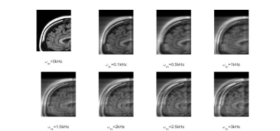
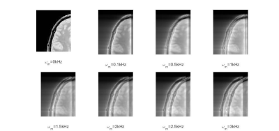
The effect of simulated source vibration at different vibration frequencies ωm on the T1 and T2 MRI image segments of human brain which lead to blurring and ghosting in the MRI image segment are illustrated in Figures 1 and 2. The brain image was acquired at 1 Tesla magnetic field and the impact of Fourier components of the vibrations on magnetic moments were artificially introduced at a computational level in order to demonstrate the impact on image quality. The vibrational effects have a relatively higher impact on overall image quality with an increase in frequency which is well reflected in the image quality.
Discussion
Specific parts of human tissues like vocal chords can vibrate at tens of KHz [6]. Other body parts for example eyes vibrate at tens of Hz [7]. The integrated effect of vibrations of body parts during MRI scan can lead to generation of a broad range of frequency components which can sum up to create blurring of images. These effects can get much more prominent in the context of MRI systems which are being developed at low magnetic fields in the range of a couple of millitesla. The overall effect of vibrations of the source to be imaged and its impact on output noise is considered a low frequency phenomenon having negligible impact on the output image which is not the case at an empirical level.Conclusion
The theoretical model and its validation through simulation proves that blurring and ghosting effects are induced in images acquired at 1T in MRI systems as a consequence of generation of side bands in the frequency band of the generated magnetic field. Our model of the noise generation process can lead to more refined algorithms in image reconstruction in MRI systems.Acknowledgements
No acknowledgement found.References
1. Emsley J.W., Feeney, J. and Emsley, J. W., Feeney, J., & Sutcliffe, L.H. High Resolution Nuclear Magnetic Resonance Spectroscopy (Elsevier, Amsterdam, 2013). Vol. 2.
2. Brown W.F., Thermal fluctuations of a single-domain particle, Phys. Rev. 1963, 130 (5), 1677.
3. Carr, H.Y. and Purcell, E.M., Effects of diffusion on free precession in nuclear magnetic resonance experiments, Phys. Rev. 1954, 94 (3), 630.
4. Woessner, D.E. Brownian motion and its effects in NMR chemical exchange and relaxation in liquids, Conc. Mag. Res. 1996, 8 (6), 397-421.
5.Pipe, J. G. Motion correction with PROPELLER MRI: application to head motion and free-breathing cardiac imaging. Magnetic resonance in medicine, 1999, 42(5), 963-969.
6. Titze, I.R. and Strong, W.J., 1975. Normal modes in vocal cord tissues. The Journal of the Acoustical Society of America, 57(3), pp.736-744.
7. Foerster, B. U., Tomasi, D. and Elisabeth C. Caparelli, E.C. Magnetic field shift due to mechanical vibration in functional magnetic resonance imaging. Magnetic Res. Med. 2005, 54 (5), 1261-1267.

