2633
Feasibility of high throughput scanning at 7T: 13 subjects per hour1Radiology, University Medical Center Utrecht, Utrecht, Netherlands, 2Philips Healthcare, Best, Netherlands
Synopsis
While substantial acceleration in MRI acquisitions have been demonstrated in the last decades (particularly at high fields where SNR is not limited), substantial patient and scan preparation time have been reported that seem to prohibit high patient throughput for clinical MRI. In this study we demonstrate that robust head MRI can be obtained at a throughput of more than 13 subjects per hour, including patient management and scan preparations (even faster than typical X-ray exams). Increased throughput may be an alternative way to “killer applications” in making high field MRI economically viable.
Introduction
The availability of MR scanners has grown enormously over the last decade. Nevertheless, MR examinations remain expensive and take a relatively long time (20-30 minutes) when compared to other imaging modalities such as X-rays, ultrasound or computed tomography. However, as MRI has a superior soft tissue contrast compared to many imaging modalities, resulting in accurate diagnostic information, and does not involve ionizing radiation, particularly important when pediatric patients are to be scanned1 reducing acquisition times, and therefore costs, can improve utilization of MRI. The need to improve utilization applies even more so when high end MRI systems are being used (like 7T), where the cost can be more than tripled compared to standard MRI systems, and its use may be more complex. As 7T MRI systems have been CE and FDA approved, the economic viability becomes an important aspect for clinical use, particularly for the diagnostic exams that do not meet with the 7T specific “killer application”. The purpose of this study was to explore the potential patient throughput in a 7T scanner for full 3D brain MRI.Methods
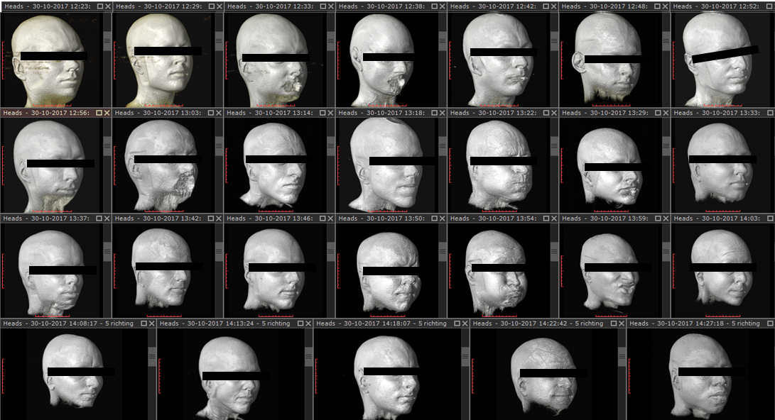
26 volunteers were scanned at a 7 tesla MR scanner (Philips, Cleveland, USA) in supine position. A head coil (NOVA medical, USA) with two transceiver channels was interfaced to two 4kW RF amplifiers. The same protocol was used for all volunteers and consisted on a 3D gradient echo, TE/TR=0.95/6 ms, FOV=250x250x250 or 275x275x250 mm3, resolution=1.5x1.5x0.75 mm3, 2m09 or 2m22 acquisition time. For visualization purposes, 3D renders were created. A large field of view was chosen to avoid the necessity of planning.Results
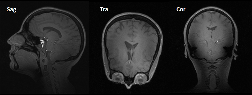
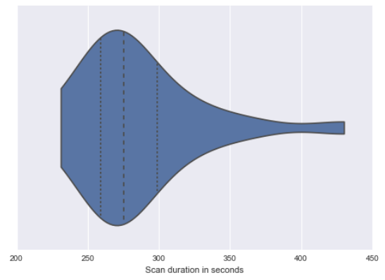
26 volunteers were successfully scanned consecutively in 2 hours and 3 minutes, leading to a mean scan time of 4m44 per person, including going in and out the scanner room. Figure 1 shows the volume rendering of the acquired images for all volunteers, demonstrating full 3D coverage of the brain. From one subject a sagittal, transverse and coronal view is shown (Figure 2). Figure 3 plots the distribution of the overall scan session times. Half of all sessions took between 4m20 and 5m00. None of the volunteers complained on the table speed used to bring the subject in or out of the isocenter of the MRI.Discussion
The results demonstrate that 26 subjects can get a 3D MRI of the brain at 7T within two hours, or a throughput of 4 minutes and 44 seconds per person. Considering an effective acquisition time of over 2 minutes, the subject handling and scan preparation can be obtained in just over 2 minutes. While our study incorporated only a single 3D scan, full clinical exams incorporate acquisitions with multiple contrasts, increasing scan time. However, highly accelerated scans have been demonstrated before using high density receiver arrays2 using SENSE3 and Caipirinha4. Further optimized acquisition schemes can even acquire multi-contrast images in around 1 minute1. Consequently when combined with under 3 minutes of preparation time, patient throughput for clinical MRI at 7T may be increased such that it becomes economically even more viable than traditional MRI.Conclusion
7T MRI can be obtained with high throughput of 13 subjects per hour for brain MRI.Acknowledgements
No acknowledgement found.References
1. Skare, S., Sprenger, T., Norbeck, O., Rydén, H., Blomberg, L., Avventi, E. and Engström, M. (2017), A 1-minute full brain MR exam using a multicontrast EPI sequence. Magn. Reson. Med.. doi:10.1002/mrm.26974
2. Petridou, N.,
Italiaander, M., van de Bank, B. L., Siero, J. C. W., Luijten, P. R. and
Klomp, D. W. J. (2013), Pushing the limits of high-resolution
functional MRI using a simple high-density multi-element coil design.
NMR Biomed., 26: 65–73. doi:10.1002/nbm.2820
3. Pruessmann, K. P., Weiger, M., Scheidegger, M. B. and Boesiger, P. (1999), SENSE: Sensitivity encoding for fast MRI. Magn. Reson. Med., 42: 952–962. doi:10.1002/(SICI)1522-2594(199911)42:5<952::AID-MRM16>3.0.CO;2-S
4. Breuer, F. A., Blaimer,
M., Mueller, M. F., Seiberlich, N., Heidemann, R. M., Griswold, M. A.
and Jakob, P. M. (2006), Controlled aliasing in volumetric parallel
imaging (2D CAIPIRINHA). Magn. Reson. Med., 55: 549–556.
doi:10.1002/mrm.20787
Figures