2618
Comprehensive analysis of advanced liver fibrosis in rats using multi-parameter MRI1Tianjin First Center Hospital, Tianjin, China, 2Radiology department, Tianjin First Center Hospital, Tianjin, China, 3Philips healthcare, Beijing China, Beijing, China
Synopsis
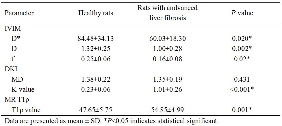
This study investigated the value of multi-parametric analysis using IVIM, DKI and MR T1ρ for the diagnosis of advanced liver fibrosis. Sixteen healthy rats and fifteen rats with advanced liver fibrosis (F4 confirmed by liver pathological examination) were scanned with IVIM, DKI and MR T1ρ. IVIM derived D*, D, f, DKI derived MD, K value and MR T1ρ derived T1ρ value were compared between the above two groups. Our results showed D*, D, f and MD decreased while K value and T1ρ value increased in rats with advanced liver fibrosis. And D*, D, f, K value and T1ρ value demonstrated significant difference (P<0.05). We therefore conclude that decreased D*, D and f and increased K value and T1ρ value could be useful in the diagnosis of liver fibrosis.
Purpose
Liver fibrosis is a common pathological change of various chronic liver diseases, endangering human health. Recent evidence suggests that liver fibrosis and even the early stage of cirrhosis can be reversible1. Diffusion dependent intravoxel incoherent motion (IVIM) and diffusion kurtosis imaging (DKI) are useful in quantifying the diffusion and perfusion effects in tissue2-4 and MR T1ρ can explore proteoglycans, collagen fibers and other macromolecular substances in tissue. The purpose of this study was to investigate the value of multi-parametric analysis using IVIM, DKI and MR T1ρ for the diagnosis of advanced liver fibrosis.Materials and Methods
Sixteen healthy rats and fifteen rats with advanced liver fibrosis (diagnosed to be with liver fibrosis F4 by liver pathological examination) were recruited. DKI, IVIM and MR T1ρ scans were acquired using a 3.0 T scanner (Ingenia, Philips, Healthcare, Best, the Netherlands) and a dStream 4-channel mouse coil. IVIM and DKI data were analyzed by using DWI post-processing software performed in a proprietary programming environment (PRIDE; Philips Medical Systems) and T1ρ map was generated on a pixel-by-pixel basis on PRIDE using a mono-exponential decay model: M(TSL)=M0*exp(-TSL/T1ρ) and T1ρ data was analyzed using the software ImageJ (available at http://rsb.info.nih.gov/ij/). Ten regions of interest (ROIs) were drawn on the upper, hilar and lower slices with attention to avoid the large blood vessels, bile ducts and artifacts. The location and size of each ROI was as same as possible in different sequences. Diffusion and perfusion related parameters (IVIM derived D*, D, f; DKI derived MD, K value;) and T1ρ value were compared by Student t test using IBM SPSS Statistics 19.0 (Armonk, New York, USA). P<0.05 indicated a significant difference.Results
MR parameters derived from IVIM and DKI and T1ρ value were compared between the two groups and summarized in table 1. Compared to control group, the perfusion related D* and the diffusion related D, f and MD decreased while K value and T1ρ value increased in rats with advanced liver fibrosis. D*, D, f, K value and T1ρ value demonstrated significant difference between the above two groups (P<0.05).Discussion
In this study, D* and f decreased in rat group with advanced liver fibrosis compared to healthy rat group, which might indicate the reduction of whole liver perfusion. The decrease of D, MD and increase of K value might result from the limitation of water diffusion, and this was consistent with the pathological changes of advanced liver fibrosis with the excessive deposition of extracellular matrix. Also the increase of T1ρ value had a great relationship with the excessive deposition of extracellular matrix. Therefore, the combination of multi-parametric MR imaging can provide more diagnostic information to give overall assessment of liver fibrosis.Conclusion
Among all parameters derived from MR IVIM, DKI and T1ρ, decreased D*, D and f and increased K value and T1ρ value could be useful in the diagnosis of advanced liver fibrosis .Acknowledgements
No acknowledgement found.References
1. Chang TT, Liaw YF, Wu SS, et al. Long-term entecavir therapy results in the reversal of fibrosis/cirrhosis and continued histological improvement in patients with chronic hepatitis B. Hepatology. 2010; 52(3): 886-893.
2. Luciani A, Viqnaud A, Cavet M, et al. Liver cirrhosis: introvoxel incoherent motion MR Imaging-Pilot study. Radiology. 2008; 249(3):891-899.
3. Goshima S, Kanematsu M, Noda Yoshifumi, et al. Diffusion kurtosis imaging to assess response to treatment in hypervascular hepatocellular carcinoma. Am J Roentgenol. 2015; 204(5):W543-549.
4. Zhao F, Wang YX, Yuan J, et al. MR T1rho as an imaging biomarker for monitoring liver injury progression and regression: an experimental study in rats with carbon tetrachloride intoxication. Eur Radiol. 2012; 22(8): 1709-1716.
Figures