2609
Robust multi-parametric mapping for abdomen imaging1Kwangwoon university, Seoul, Republic of Korea
Synopsis
A robust abdominal multi-parametric mapping using multi-echo data is proposed. Reconstructed maps are water, fat images, quantitative susceptibility map (QSM), and R2* map. Fat fraction and iron deposition in the liver may be important parameters for diagnosis. Challenges to the abdominal mapping include large field inhomogeneity, phase wrapping, phase variations from water and fat signal, chemical shift, and physiological motions. We applied simultaneous unwrapping phase and error recovery from inhomogeneity (SUPER) technique to correct field inhomogeneity and phase wrapping. The technique is stably applicable to objects containing water and fat signal, and is also useful as a preprocessing for QSM.
Introduction
Multi-parametric mapping for abdomen has several challenges including large field inhomogeneity, phase wrapping, phase variations from water and fat signal, chemical shift, and physiological motions.1 Fat saturation may be useful to remove fat-related phase variation, however it requires high field homogeneity, which is hardly achievable in abdomen imaging or mapping.2 A long r.f. pulse for fat saturation is also an obstacle to GRE imaging with a short TR for a breath hold. In addition, fat is useful information in some organs such as liver. Thus the approach should maintain water and fat information, and correct inhomogeneity from water and fat phases.3 Phase unwrapping is also needed to estimate inhomogeneity properly.Methods
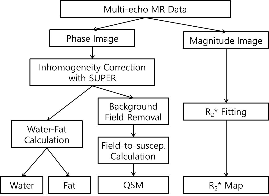
Three-dimensional (3-D) multi-echo data is first acquired. Simultaneous unwrapping phase and error recovery from inhomogeneity (SUPER) method is applied to correct inhomogeneity.4 SUPER corrects field inhomogeneity, and unwraps phase simultaneously, by which water and fat images are accurately separated. SUPER models field inhomogeneity with a polynomial, and finds model coefficients from the partial derivatives of the model formula using measured phase difference data. Both water and fat phases are used. Correction of the global inhomogeneity is useful for QSM, to remove the phase not satisfying the Laplace equation. After correction of the phase, QSM is obtained by the field-to-susceptibility conversion formula. R2* map is obtained from the multi-echo magnitude images. Figure 1 depicts a flow chart of the proposed method.Results
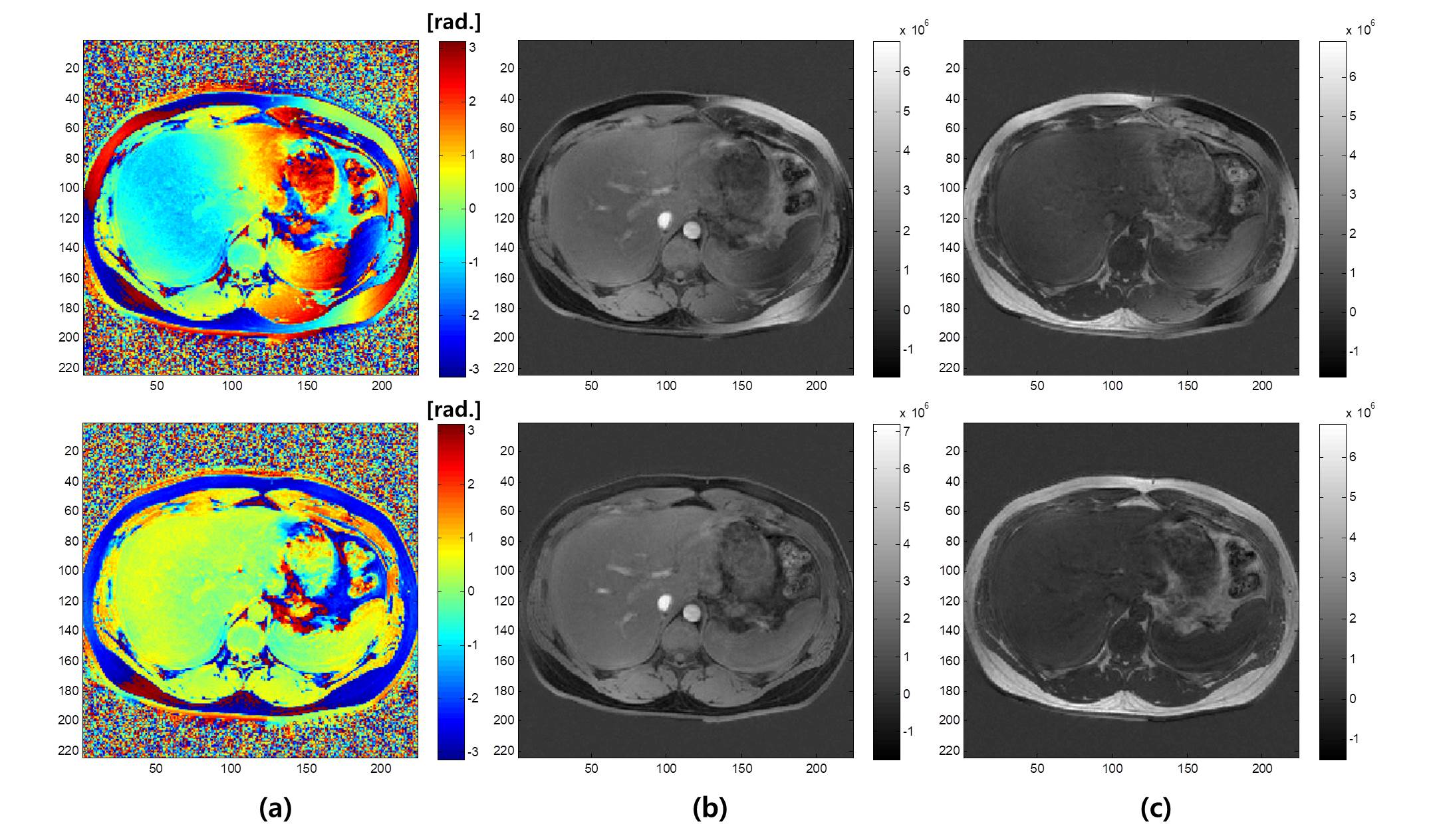
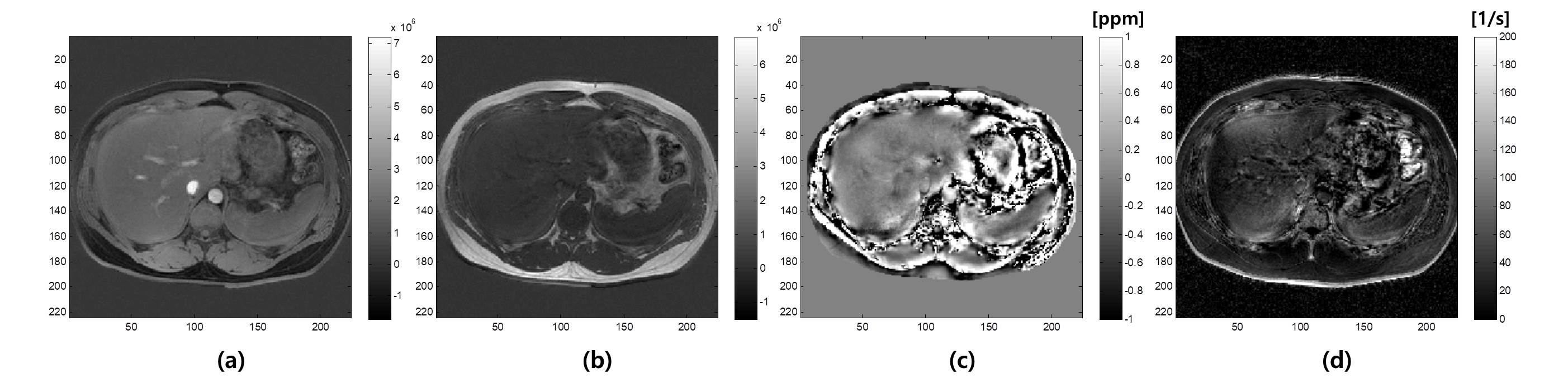
3-D GRE sequence was used at 3.0 Tesla MRI system (Philips, Achieva) for in-vivo abdominal imaging. Eight echoes were acquired with parameters: TR/TE1/DTE = 15/1.15/1.7ms, voxel resolution = 1.8/1.8/3.0 mm, BW/pixel = 1215Hz, flip angle = 15 degrees, number of channels = 16, matrix size = 224x224. Figure 2 shows a sample phase map of the out-of-phase image (a), water-only (b), and fat-only (c) images. The images at the top are before correcting field inhomogeneity, and those at the bottom are after correcting it by SUPER. Figure 3 shows multi-parametric maps for abdomen by the proposed method: (a) water-only, (b) fat-only, (c) susceptibility map, and (d)R2* map.Discussion
Abdominal multi-parametric mapping (fat/water, QSM, and R2*) is proposed with multi-echo data for quantitative imaging. Fat fraction is a useful parameter in abdominal imaging. QSM and R2* mapping are also useful to evaluate iron deposition in the liver, which is a good indicator of the iron concentration in the body.5 We applied SUPER method to correct global field inhomogeneity, by which improved water/fat separation and QSM are obtained. SUPER can remove some global phase not satisfying the Laplace equation, which may not be removable by conventional background phase removal method.Conclusions
Using the proposed method, abdominal multi-parametric mapping was successfully obtained in a single breath-hold. Water-fat images, QSM, and R2* were obtained from 8 echoes, which are useful for diagnosis. Using SUPER, field inhomogeneity was properly corrected, by which improved water-fat images and QSM, R2* map were obtained.Acknowledgements
This work was supported by the National Research Foundation of Korea (NRF) grant funded by the Korea government (MSIP) (NRF-2015R1A2A2A03005089). The present research has also been conducted by the research grant of Kwangwoon University in 2017.References
1. Dong J, Liu T, Chen F, Zhou D, Dimov A, Raj A, Cheng Q, Spincemaille P, Wang Y. Simultaneous phase unwrapping and removal of chemical shift (SPURS) using graph cuts: Application in quantitative susceptibility mapping. IEEE Trans Med Imaging 2015;34(2):531-40.
2. Delfaut EM, Beltran J, Johnson G, Rousseau J, Marchandise X, Cotten A. Fat suppression in MR imaging: Techniques and pitfalls. Radiographics 1999;19(2):373-82.
3. Reeder SB, McKenzie CA, Pineda AR, Yu H, Shimakawa A, Brau AC, Hargreaves BA, Gold GE, Brittain JH. Water–fat separation with IDEAL gradient‐echo imaging. Journal of Magnetic Resonance Imaging 2007;25(3):644-52.
4. Yang Y, Park J, Yoon J, Ahn C. Field inhomogeneity correction using partial differential phases in magnetic resonance imaging. Phys Med Biol 2015;60(10):4075.
5. Brittenham GM, Badman DG, National Institute of Diabetes and Digestive and Kidney Diseases (NIDDK)Workshop. Noninvasive measurement of iron: Report of an NIDDK workshop. Blood 2003;101(1):15-9.
Figures