2606
Hepatic Iron overload estimation by proton density mDIXON Quant technique1Donostia Hospital, San Sebastian, Spain, 2Philips Healthcare Iberia, Madrid, Spain, 3Osatek, San Sebastian, Spain, 4Basque Country University, San Sebastian, Spain
Synopsis
This work evaluates the utility of R2* obtained from multi-point multi-peak proton density fat fraction to assess iron overload and the accuracy of provided relaxation maps compared with more established multi-echo gradient echo sequence.
Introduction
Multi-point multi-peak DIXON techniques have shown high reproducibility to estimate proton density fat fraction among different magnetic fields and different imaging platforms (1). These methods take into account R2* exponential decay to prevent R2* relaxation effects on fat fraction estimation (2,3). The resulting R2* maps can be used to assess iron overload in combination with fat fraction infiltration in a single acquisition. This work evaluates the utility of R2* obtained from multi-point multi-peak proton density fat fraction to assess iron overload and the accuracy of this relaxation map compared with more established multi-echo gradient echo sequence.Material and Methods
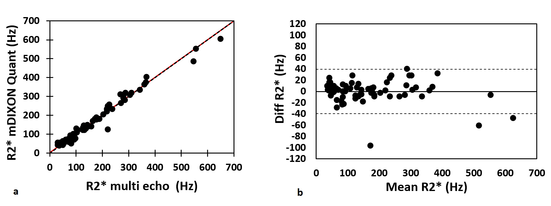
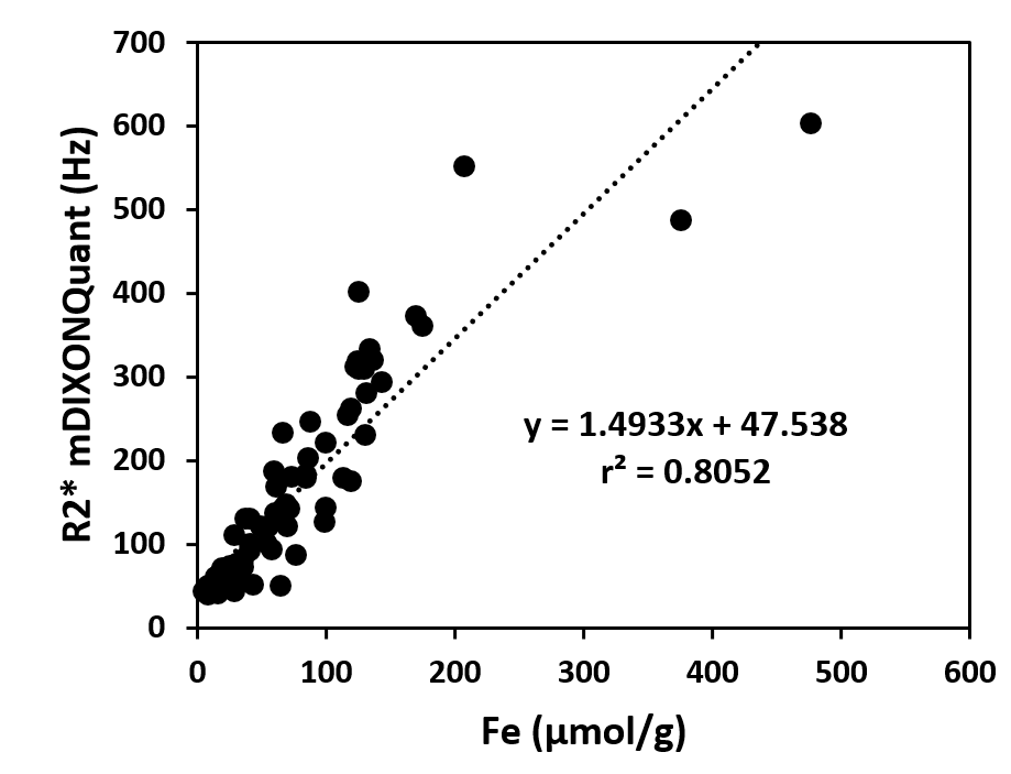
The research project was approved by the Institutional Research Board and all participant signed informed consent. Seventy-two patients with different degrees of iron overload were enrolled in the study. All acquisitions were acquired in a 1.5T Ingenia system (Philips Heathtech, Best, The Netherlands) using a phased array Torso/Cardiac coil with 28 elements. The magnetic resonance protocol included: The approved FerriScan (Resonance Health, Burswood, Australia) protocol for non-invasive estimation of iron content in the liver in all patients, a mDixon acquisition and a 3D multi-echo gradient echo sequence. Six echoes mDIXON Quant acquisition (TR/Initial TE/∆TE/flip angle = 6.5ms\1.16ms\0.8ms\5o) was used to obtain water, fat, fat fraction and T2* maps using accurate seven-peaks fat model and eddy current compensation. 3D multi-echo gradient echo sequence (TR/Initial TE/∆TE/flip angle= 0.91ms/0.82ms/5o) with 20 echoes was acquired as gold standard to validate the T2* values obtained from the mDIXON acquisition. T2* maps were generated at the scanner using a nonlinear maximum likelihood expectation maximization algorithm to take into account not Gaussian noise distribution in those images with very low signal. R2* values were obtained in 3 regions of interest (ROIs) placed in right liver lobe avoiding big vessels. Equivalent ROIs were placed for both R2* maps obtained from mDIXON quant and multi-echo gradient echo sequence. Final reported values were obtained as the average of the three different regions. Same ROIs were also used to estimate fat fraction content. Passing Bablok regression analysis and Bland-Altman plots were performed to compare R2* obtained by both techniques. The R2* values obtained from mDIXON Quant sequence were compared with the iron content obtained from FerriScan methodology.Results and Discussion
Iron concentration in the cohort ranged from 5 μmol Fe/g to 477 μmol Fe/g (78.63±76.30 μmol Fe/g). Fat Fraction content ranged from 3.6% to 35.9%. Passing-Bablok regression analysis obtained a slope of equal to 0.995 (0.96-1.03, 95% CI) and intercept equal to 2.83 (-2.22,6.55, 95% CI) (Figure 1a). Bland-Altman plot between both R2* values presents minimal bias (mean difference -0.33Hz) with none significant trend with the increment of R2* values (Figure 1b). MDIXON Quant R2* values correlate well with iron content obtained from the FerriScan methodology (r2=0.80) although R2* values above 210 μmol Fe/g present limited increment on R2* values (Figure 2).Conclusions
R2* values obtained from mDIXON Quant technique show good correlation with Iron overload, although R2* values present some saturation for iron overloads higher than 210 μmol Fe/g. In addition, R2* values obtained by mDIXON Quant show an excellent correlation with R2* values acquired with a standard multi-echo gradient echo with higher number of echoes. In conclusion R2* obtained for mDIXON Quant acquisition present equivalent accuracy than conventional R2* multi-echo acquisition and good sensitivity to estimate liver iron overload.Acknowledgements
No acknowledgement found.References
1. Serai SD, Dillman JR, Trout AT. Proton Density Fat Fraction Measurements at 1.5- and 3-T Hepatic MR Imaging: Same-Day Agreement among Readers and across Two Imager Manufacturers. Radiology. 2017 Jul;284(1):244-254.
2. Yu H, Shimakawa A, McKenzie CA, Brodsky E, Brittain JH, Reeder SB. Multiecho water-fat separation and simultaneous R2* estimation with multifrequency fat spectrum modeling. Magn Reson Med. 2008 Nov;60(5):1122-34.
3. Eggers H1, Brendel B, Duijndam A, Herigault G. Dual-echo Dixon imaging with flexible choice of echo times. Magn Reson Med. 2011 Jan;65(1):96-107.
Figures