2605
Quantification of multiple organ iron deposition in transfusion dependent diseases using mDIXON-Quant technique1Tianjin University of Traditional Chinese Medicine, Tianjin, China, 2Clincial Science, Philips Healthcare, Beijing, China, 3Tianjin First Center Hospital, Tianjin, China
Synopsis
Iron overload is a common complication of transfusion dependent patients. Magnetic resonance imaging can be used for quantitative detection of iron deposition in transfusion dependent patients. A total intake of iron for transfusion was evaluated based on the mDIXON-Quant and 3D-FFE sequence respectively. Because the mdixon-quant can avoid the effect of fat on the iron overload evaluation, the mDIXON-Quant sequence can more accurately quantify iron deposition in liver and pancreas than 3D-FFE sequence. The quantitative application of mDIXON-Quant in detection of iron deposition in patients can provide reliable basis for iron chelation therapy in clinic.
Introduction
Transfusion dependent disease is generally caused by long-term repeated blood transfusions during clinical treatments, which was usually found in beta thalassemia, aplastic anemia (AA) and sickle cell anemia disease(SCD). Iron overload is one of the common complications and could furtherly lead to different degrees of tissue degeneration, necrosis and fibrosis. In this work, we tried to investigate the clinical value of mDIXON-Quant in the quantitative evaluation of transfusion dependent patients with multiple organ iron deposition, which would have a very important role in the evaluation of organ function and guiding for clinical chelated iron treatment plan.Methods
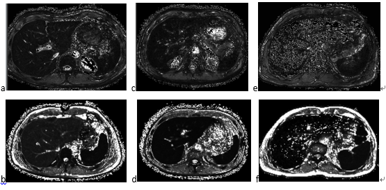
22 transfusion dependent patients who haven’t undergone iron chelation therapy were recruited in this study. This study was approved by the ethics committee of Tianjin First Centre Hospital, Tianjin, China. And all the subjects or their families were informed of the purpose of the study. According to the volume of blood transfusion, the subjects were divided into non transfusion group (10 cases without using blood products containing red cells for 3 years), small amount of blood transfusion group (5 cases with blood transfusion of 0-20u within 3 years, same as an Yu Tie intake dose of 1~63.2mg/kg) and massive blood transfusion group (7 cases with blood transfusion of greater than 20u within 3 years, same as an Yu Tie intake dose of 63.2mg/kg). All patients underwent MR examinations based on a Philips Ingenia 3.0T MR scanner with multi-echo 3D-FFE and 3D mDIXON-Quant protocols. The scanning parameters of 3D multi-echo FFE were as follows: TR=65ms, TE=3.0ms, 6.4ms, 9.8ms, 13.2ms and 16.6ms, slice thickness=4mm, FOV=320x280x12mm3, voxel size=2x2x4mm3. mDIXON-Quant scanning parameters were as follows: TR=6.9ms, TE=1.06ms, 1.96ms, 2.86ms, 3.76ms, 4.66ms and 5.56ms, slice thickness=4mm, FOV=320x280x12mm3, voxel size=2x2x4mm3. For 3D multi-echo FFE images, the R2* (R2*=1/T2*) of liver and pancreas were calculated voxel by voxel. For mDIXON-Quant images, the dR2* (dR2*=1/dT2*), which for distinguishing from the R2* with 3D multi-echo FFE, were calculated within liver and pancreas voxel by voxel. Pearson’s correlation was performed to investigate the relationship of the R2* (and dR2*) of liver and pancreas with the cumulative intake of iron during blood transfusion.Results
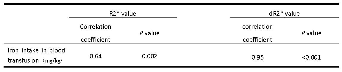
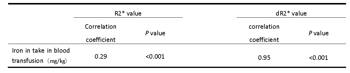
The results showed that liver R2* and dR2* value were significantly correlated with total transfusion iron intake with the correlation coefficient of 0.64(P=0.002) and 0.95(P<0.001) respectively. There was week correlation between the pancreatic R2* and the total iron intake with the correlation coefficient of 0.29(P<0.001). But there is strong correlation between pancreatic dR2* and total volume of blood transfusion with the correlation coefficient of 0.95 (P<0.001).Discussion
The results showed that both multi-echo 3D FFE and mDIXON-Quant imaging can be applied to reflect the iron deposition different organs. mDIXON-Quant technique is based on the proton chemical shifts of fat and water with a multi-echo data acquisition. Compared to multi-echo 3D-FFE sequence, mDIXOn-Quant can avoid the interference of adipose tissue, which would be better for the quantitative evaluation of transfusion dependent patients with multiple organ iron deposition.Conclusion
Both multi-echo 3D FFE and mDIXON-Quant can be applied for the evaluation of organ iron deposition, which would be helpful for the clinical diagnosis and therapy evaluation.Acknowledgements
No acknowledgement found.References
[1] Wahidiyat PA, Liauw F, Sekarsari D. Evaluation of cardiac and hepatic iron overload in thalassemia major patients with T2* magnetic resonance imaging. Hematology. 2017 Feb 20:1-7.
[2] Ramanathan.Ganesh,Olunyk.John K. Diagnosing and preventing iron overload.[J].2017,21:S58-S67.
[3] KARIMI Z M H, G H A S M H N M B M. Serum Ferritin Levels Correlation With Heart and Liver MRI and LIC in Patients With Transfusion-Dependent Thalassemia[J]. Iranian Red Crescent medical journal, 2015, 17(4): 1–4.
Figures