2586
Long-term and short-term repeatability of hepatic proton density fat fraction measurement across MR field strengths in nonalcoholic fatty liver disease subjects and a phantom1Radiology, Ajou University Hospital, Suwon, Republic of Korea, 2Internal Medicine, Ajou University Hospital, Suwon, Republic of Korea
Synopsis
Long-term and short-term repeatability of hepatic proton density fat fraction measurements was assessed across MR field strengths in nonalcoholic fatty liver disease subjects and a phantom. Our results showed that PDFF measurement have high short-term and long-term repeatability across the fields strengths, and patients undergoing a longitudinal PDFF measurement may be scanned regardless of MR field strength.
Introduction
MR-based proton density fat fraction (PDFF) measurements have shown high sensitivity and specificity in detecting and quantifying fat deposition in the liver (1-4). Given its noninvasiveness and accuracy in hepatic steatosis quantification, PDFF has strong advantages in long-term monitoring of steatotic patients and response to treatment. In order to determine the significance of fat fraction (FF) changes during a longitudinal follow-up, repeatability of the PDFF measurements needs to be established beforehand. The purpose of our study is to assess the long-term and short-term repeatability of PDFF measurements across MR field strengths in nonalcoholic fatty liver disease (NAFLD) subjects and a phantom.Methods
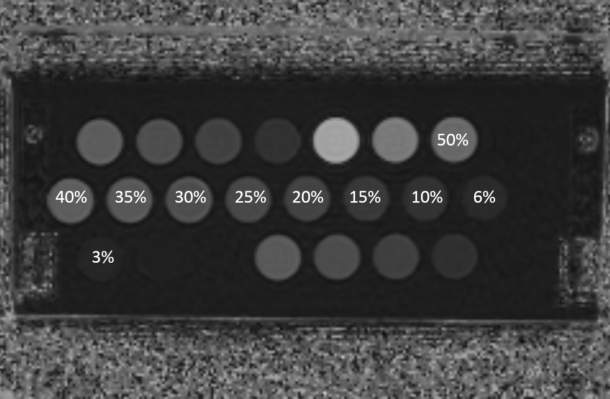
A phantom consisted of 10 test tubes containing 3%-50% of lard was scanned on 1.5 T (Signa HDxt; GE) and 3.0 T (Discovery 750 W; GE) units within a day and a 14 days interval, with five repeated PDFF measurements per each scan in addition to MR spectroscopy (Figure 1). For clinical scan, fourteen prospective-enrolled NAFLD patients (9 males, 5 females; average ± SD of age, 45.8 years ± 15.3; age range, 19-72 years) were referred from the Liver Center at our institution. They underwent PDFF scans on 1.5 T and 3.0 T units on the same day, within a 15 minute interval, followed by identical scans on 10-14 days (mean, 12.8 days; median, 14 days) after. Each MR scan consisted of two consecutive PDFF measurements, resulting in a total of four PDFF scans per day. A commercially available PDFF pulse sequence (IDEAL IQ, GE) of low flip angle (3˚-5˚), six echoes, and multipeak fat model was used for all scans. To estimate hepatic PDFF, free-hand region of interests were drawn on each Couinaud segment using a vendor-provided software (READY View, GE). All subjects were asked to refrain from consuming excessive fatty meal or alcohol during the recess. The repeatability of PDFF measurements in terms of interscan interval and field strengths in human and the phantom was assessed by within-subject coefficient of variation (wCV). The correlation of PDFF measurements between field strengths was assessed. Also, the bias between the MRS- and PDFF-measured FF across field strengths was evaluated in the phantom.Results
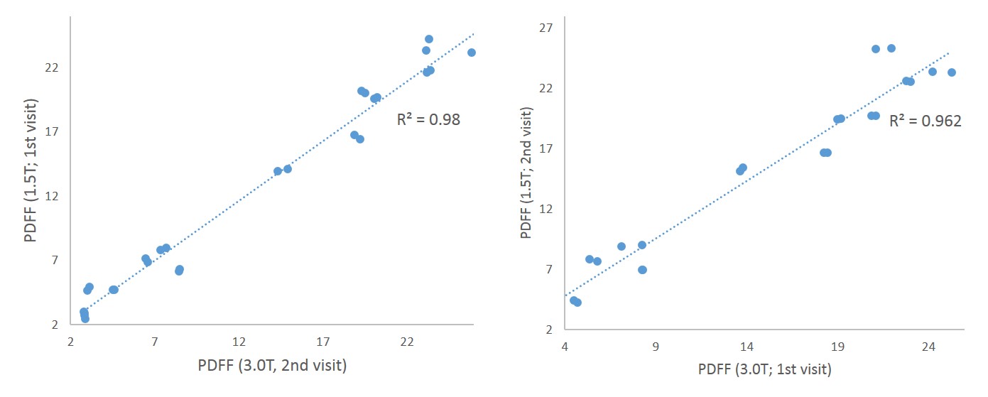
In the phantom scan, MRS- and PDFF-measured FF showed good correlation in both 1.5 T and 3.0 T scanners (r2=0.906, 0.927, respectively). PDFF-measured FF across field strengths showed strong correlation (r2=0.996) with mean difference of 1.07% and 95% limits of agreement ranging ± 0.14 on Bland-Altman analysis. Within-day and two-week wCV was 0.28%-0.64% and 1.56% for 1.5 T, 0.13%-0.27% and 2% for 3.0 T, and 0.26%-5.9% and 4.2%-4.7% across field strengths. In human subjects, the mean ± SD of the subjects’ BMI and PDFF at 3.0T was 28.0 kg/m2 ± 5.4 and 12.7% ± 8.4 (range, 2.72%–35.3%), respectively. BMI and body weights of the subjects showed moderate positive correlation with the PDFF measurements (r2=0.574, 0.552, respectively). PDFF-measured FF across field strengths showed strong correlation, with r2=0.990 and 0.993 for the same day, and 0.962 and 0.98 for two weeks interval (Figure 2). Within-day and two-week wCV was 0.98%-1.3% and 6.3% for 1.5 T, 2.1%-3.2% and 6.4% for 3.0 T, and 3.6%-4.5% and 6.1%-7.5% across field strengths. PDFF measurements exclusively from right and left livers showed 7.8% and 8.2% of wCV across field strengths.Discussion and Conclusion
Our results indicated that PDFF measurement have high short-term and long-term repeatability across the fields strengths. In both phantom and clinical scan, within-day repeatability in the same MR field strength was excellent, with 1.5 T showing better results than 3.0 T. Long-term repeatability across field strengths in clinical scan was comparable to that of the same field strength with wCV of 6.1%-7.5%. Although repeatability of PDFF measurement was better in the right liver than in the left liver, it was still poor than measuring the whole liver segments. In conclusion, repeatability of PDFF measurement across field strengths is comparable to that of the same field strength, and patients undergoing a longitudinal PDFF measurement may be scanned regardless of MR field strength.Acknowledgements
No acknowledgement found.References
1. Middleton MS, Heba ER, Hooker CA, Bashir MR, Fowler KJ, Sandrasegaran K, et al. Agreement Between Magnetic Resonance Imaging Proton Density Fat Fraction Measurements and Pathologist-Assigned Steatosis Grades of Liver Biopsies From Adults With Nonalcoholic Steatohepatitis. Gastroenterology 2017;153:753-761
2. Tang A, Tan J, Sun M, Hamilton G, Bydder M, Wolfson T, et al. Nonalcoholic fatty liver disease: MR imaging of liver proton density fat fraction to assess hepatic steatosis. Radiology 2013;267:422-431
3. Idilman IS, Aniktar H, Idilman R, Kabacam G, Savas B, Elhan A, et al. Hepatic steatosis: quantification by proton density fat fraction with MR imaging versus liver biopsy. Radiology 2013;267:767-775
4. Kuhn JP, Hernando D, Munoz del Rio A, Evert M, Kannengiesser S, Volzke H, et al. Effect of multipeak spectral modeling of fat for liver iron and fat quantification: correlation of biopsy with MR imaging results. Radiology 2012;265:133-142
Figures