2585
Measurement of Hepatic Lipid During Free Breathing with T2-Corrected Multiecho 1H MR SpectroscopyJack Knight-Scott1, Adina Alazraki1,2, Miriam Vos2, Xiaodong Zhong3, and Brian Dale4
1Radiology, Children's Healthcare of Atlanta, Atlanta, GA, United States, 2Pediatrics, Emory University, Atlanta, GA, United States, 3MR R&D Collaborations, Siemens Healthineers, Los Angeles, CA, United States, 4MR R&D Collaborations, Siemens Healthineers, Cary, NC, United States
Synopsis
Current MR techniques for quantifying hepatic fat through measurement of the proton density fat fraction (PDFF) require a breath hold that many patients find challenging. In this work, we show that when employing single voxel multiecho spectroscopy for measurement of the liver PDFF, breath holding and free breathing acquisitions yield similar results.
Introduction
Magnetic resonance imaging (MRI) and spectroscopy (MRS) have gained increasing favor in recent years as non-invasive techniques for measuring and monitoring hepatic fat in non-alcoholic fatty liver disease (NAFLD1,2). One such technique, high-speed T2-corrected multi-echo 1H spectroscopy (HISTO3), employs a specialized stimulated echo acquisition mode spectroscopy localization sequence to acquire data at multiple echo times within a single breath hold. When combined with a moderate repetition time (TR), HISTO allows measurement of the proton density fat fraction (PDFF) with minimal relaxation weighting. Because current MR liver fat techniques require a breath hold – HISTO included – these techniques have limited application in patients where breath holding might be particularly challenging, such as with the elderly and children. While the effects of respiratory motion in abdominal imaging are well understood, its effects in abdominal spectroscopy have been little studied. The purpose of this study was to evaluate the efficacy of HISTO during free breathing.Methods
All studies were approved by the IRB and performed on a 1.5 T MRI system (Siemens Healthineers, Erlangen, Germany) using a 16-channel body array coil in conjunction with a spine coil. Breath-hold (BH) and free-breathing (FB) liver spectra were acquired from 18 participants (10/8 female/male, age range: 9-19 yrs, mean: 15.8±2.3 yrs) using a commercial HISTO sequence. Repeatability data was collected on 7 of the participants (3/4 female/male): participants were removed from the magnet, taken off the table, placed back on the table and repositioned for the second set of measurements. Voxels were placed away from major vessels, the left liver lobe, and the edges of the liver. No attempt was made to position the voxel in the same region for repeat measurements. The HISTO sequence was run with a 3000-ms TR, a 10-ms mixing time (TM), and five echo times: 12, 24, 36, 48 and 72-ms (Figure 1). Signals consisted of 1024 complex points acquired with a 1200 Hz spectral width from a 3x3x3 cm3 voxel. Liver fat data sets were acquired during a 15 s free-breathing period, then repeated for a 15 s breath hold period. Liver PDFFs were obtained from automatically generated HISTO reports. Water line widths were measured using AMARES in jMRUI4,5. The relation between BH and FB liver PDFF was examined through linear regression. To examine repeatability, we employed linear regression and measured differences in PDFF in the sub group of 7 participants for each FB-BH pair, and for the first and second set of FB data, and first and second set of BH data.Results
Agreement between BH and FB liver fat measurements is excellent (Figure 2) with a coefficient of determination r2 of 0.997, and a regression curve with a slope of 0.997 (p < 0.00001) and an intercept of only 0.095. For repeated measurements, all r2 values were greater than 0.990 s (Table 1). Differences between the BH and FB fat measurements for all 25 data sets ranged from -1.32 to 0.96 percentage points. Surprisingly, but not significantly (p=0.306, N=18, Signed Rank Test), line widths were in general wider for BH than FB measurements. Correlation analysis of all repeated measurements yields values greater than 0.995 for all possible combinations, an indication that differences between repeated sets are not significant.Discussion
With careful voxel placement, a 15 s HISTO sequence can be performed without a breath hold. This makes the sequence very useful in pediatrics, sedated patients, and others with breathing and respiratory challenges. Furthermore, these results indicate that multiple liver regions can be spectroscopically interrogated without additional stress to patients. Although differences between successive FB and BH measurements are on average less than one percentage point, the largest difference occurs when the BH spectral line width is more than 100% greater than that of the FB line width. Visual verification of spectral quality might thus be required for longitudinal measurements when tracking small changes – less than 2-3 percentage points. Current results strongly support free-breathing spectroscopic liver PDFF measurements during multi-echo acquisitions.Acknowledgements
Thanks to all the MR Technologists at Children's for their help with this study.References
- Springe F, Machann J, Claussen CD et al, Liver fat content determined by magnetic resonance imaging and spectroscopy. World J Gastroenterol. 2010;16(13):1560-1566.
- Reeder SB, Cruite I, Hamilton G et al. Quantitative assessment of liver with magnetic resonance imaging and spectroscopy. J Magn Reson Imaging. 2011;34:729-749.
- Pineda N, Sharma P, Xu Q, et al, Measurement of hepatic lipid: high speed T2-corrected multiecho acquisition at 1H MR spectroscopy – a rapid and accurate technique. Radiology. 2009;252(2):568-576.
- Vanhamme L, van den Boogaart A, Van Huffel S. Improved method for accurate and efficient quantification of MRS data with use of prior knowledge. J Magn Reson. 1997;129:35-43.
- Naressi A, Couturier C, Devos JM, et al. Java-based graphical user interface for the MRUI quantitation package. Magma. 2001;12(2-3):141–152.
- Stefan D, Cesare FD, Andrasescu A, et al. Quantitation of magnetic resonance spectroscopy signals: the jMRUI software package. Meas Sci Technol. 2009;20(10):104035.
Figures
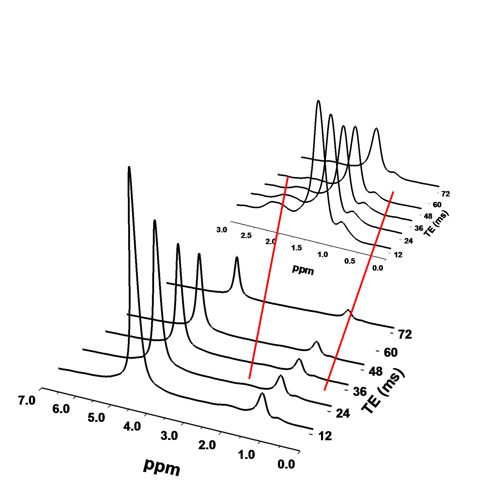
Figure 1. Representative
HISTO Liver Spectra. Water and lipids peaks as a function of TE: 12, 24, 36, 48 and 72 ms. A zoomed portion shows lipid peaks within 0 to 3.0
ppm region. Each
spectrum was acquired with 1 NEX, 10 ms TM, and a 3000 ms TR.
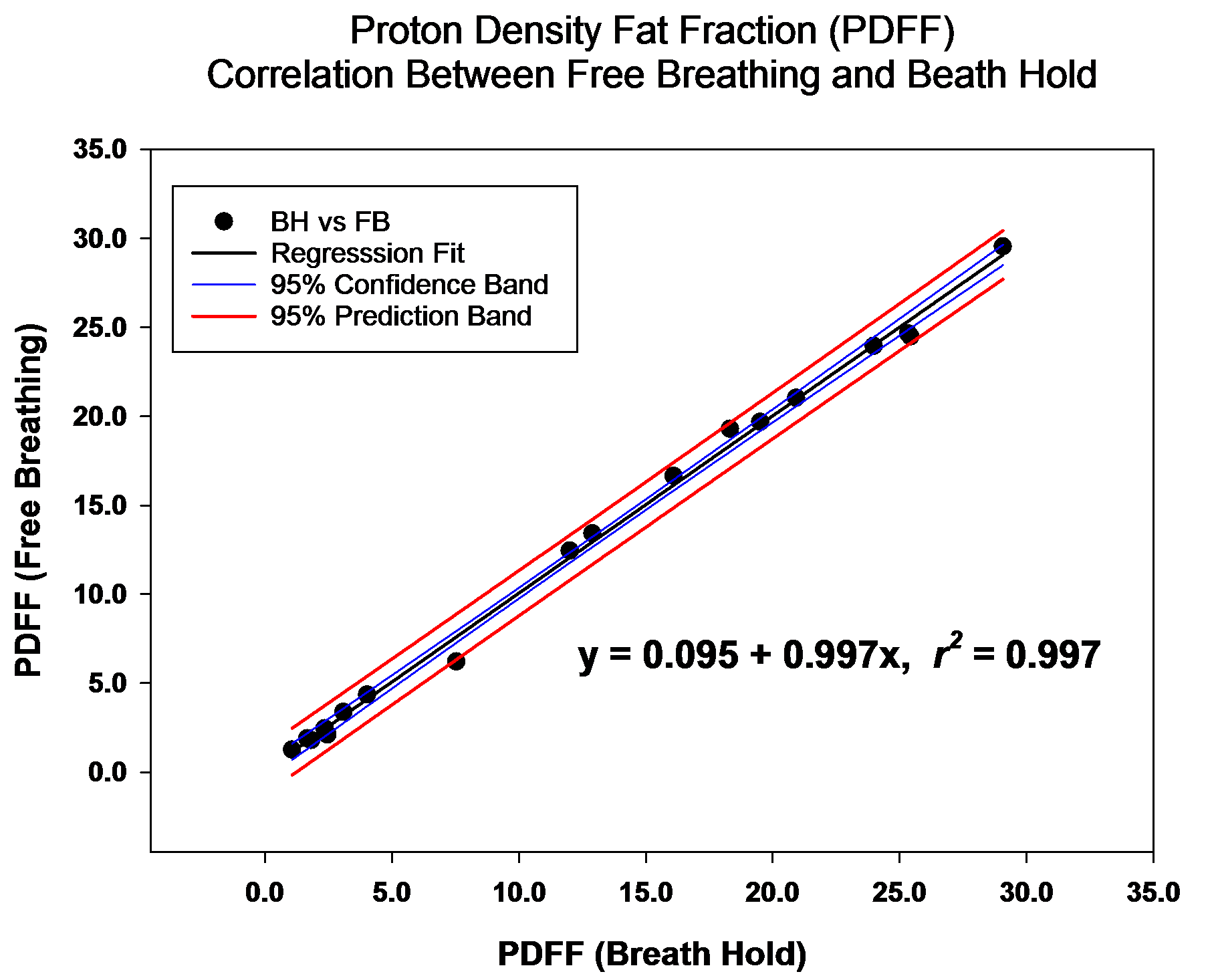
Figure 2. Regression
of Free-Breathing Liver PDFF vs Breath-Hold Liver PDFF (N=18).
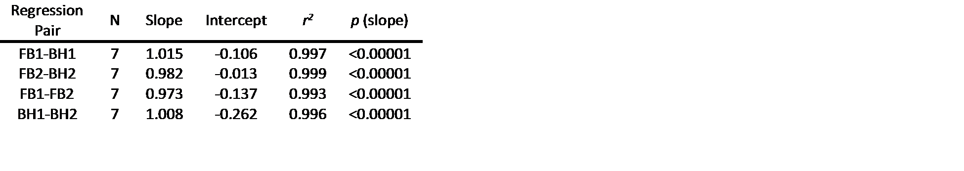
Table 1. Linear Regression Results for Repeatability. Slope, intercept, r2, and p-value for the
slope.