2572
Motion-tolerant super-resolution reconstruction from multi-stack MR data1Radiology, Columbia University Medical Center, New York, NY, United States, 2Radiology, New York Presbyterian Hospital, New York, NY, United States, 3Global MR Applications and Workflow, GE Healthcare, New York, NY, United States, 4Biomedical Informatics, Columbia University, New York, NY, United States, 5Radiology, Columbia University Medical Center, NY, NY, United States, 6Radiology, New York Presbyterian Hospital, NY, NY, United States, 7New York State Psychiatric Institute, New York, NY, United States, 8Psychiatry, Columbia University, 10032, NY, United States
Synopsis
Image super resolution reconstruction (ISRR) is a technique that may be useful for generating fast, motion tolerant 3D reconstructed images from multi stack data. We provide initial results of a multi-step ISRR approach using patch-to-volume reconstruction(PVR) followed by a slice-by-slice convolutional neural network to further improve spatial resolution. Our methods provide improved measures of peak-SNR, and could be used to rapidly generate 3D volumes from multiple 2D stacks in fetal and abdominal imaging where constant motion requires short scan times as well as in pelvic imaging where high SNR requirements lead to long scan times and motion artifact. Motion artifact is a significant obstacle in these MRI applications resulting in image quality degradation and potentially limited diagnostic ability.
Purpose/Introduction
Many routine clinical MRI protocols require multi-plane T2-weighted views or high-resolution 3D T2 acquisition, as in prostate cancer diagnosis and biopsy targeting, for example. These acquisitions tend to be time-consuming, ranging from from 3 to 5 minutes for each axial, coronal and sagittal 2D sequence and longer for a single volumetric 3D sequence. The long scan times often result in motion artifact which degrade image quality, hamper diagnosis and further prolong overall scanner time. A possible strategy to reduce acquisition time and therefore reduce motion artifact is to acquire fast multiple stacks of low resolution sequences and use ISRR techniques for reconstruction of higher resolution images.Methods
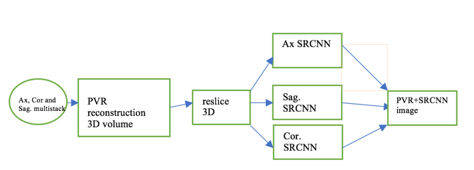
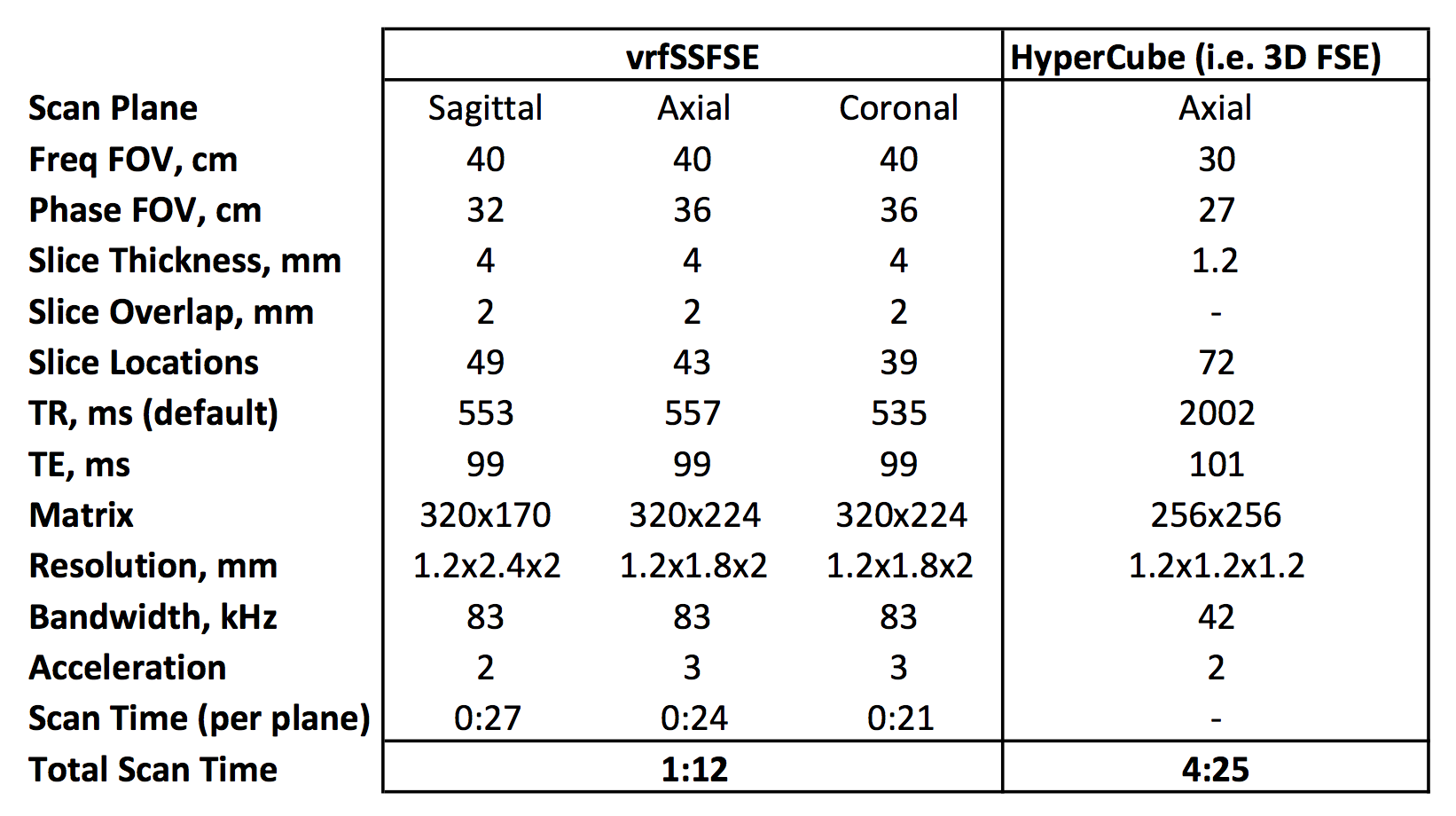
Data acquisition: Two volunteers who provided informed consent were scanned on a 60-cm 3T MRI scanner (Discovery MR750, Waukesha, WI) using a 32-channel phased array. Single-shot fast spin echo imaging with variable refocusing flip angles (vrfSSFSE) was performed in three orthogonal planes with similar scan parameters (Table 1). The stacks were acquired with low through-plane resolution (LR) of 4 mm slice thickness with 50% overlap between adjacent slices. For comparison, a high-resolution (HR) axial 3D FSE isotropic volume scan was also acquired with T2 weighting. For SR 3D volume, a 2-step reconstruction process was followed. For motion-insensitive SR reconstruction, first patch-to-volume reconstruction (PVR) algorithm [1] was applied to generating a 3D volume. This was followed by applying super resolution convolution neural network (SRCNN) [2] for each slice. Three SRCNN models were created from axial, coronal and sagittal with high in-plane resolution SSFSE images for training the SRCNNs as shown in Figure 1. For each CNN model, training data was prepared by using 32x32 overlapping patches from each SSFSE images along with a low-resolution version of those patches. For testing, the 3D volume from PVR was resliced into 3 orthogonal stacks and slice-by-slice SR trained model was applied. The SRCNNs were implemented using Keras (https://github.com/fchollet/keras). The three multi-slice SR stacks were averaged together to generate SRCNN based volume. For comparison 3D FSE was used as the reference standard image. Peak signal-to-noise ratio (PSNR) was used to evaluate the quality of reconstructed SR image quantitatively. PSNR is defined as, $$PSNR = 10.\log_{10}{\left(\frac{MAX_I^2}{MSE} \right)}$$ where MSE is the mean square error between the HR 3D FSE image and the generated super resolution image, MAXI is maximum possible pixel value in HR image.
Results
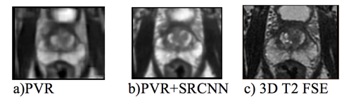
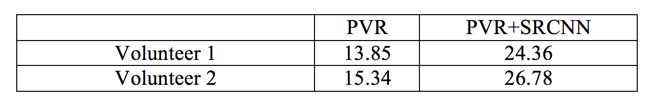
Super Resolution reconstruction of three LR image stacks resulted in a single HR volume. In Figure 2, the image on the left displays the PVR image in axial view, the middle shows the PVR+SRC NN reconstructed HR image and the gold standard 3D FSE HR volume is shown on the right. The results for PSNR for the cases are shown in Table 2.Discussion/Conclusion
We have presented a method for reconstructing an HR volume from multiple LR stacks. This SR method could be used for rapid 3D imaging and/or applications where motion artifacts tend to be problematic, such as fetal abdominal imaging etc. In the future, we plan to evaluate reconstructed HR volume under clinical conditions and as a function of number of stacks and native stack resolution.
Acknowledgements
No acknowledgement found.References
1) Alansary A, Rajchl M, McDonagh SG, Murgasova M, Damodaram M, Lloyd DFA, Davidson A, Rutherford M, Hajnal JV, Rueckert D, Kainz B. PVR: Patch-to-Volume Reconstruction for Large Area Motion Correction of Fetal MRI. IEEE Trans Med Imaging. 2017 Oct;36(10):2031-2044. doi: 10.1109/TMI.2017.2737081. Epub 2017 Sep
2) Chao Dong, Chen Change Loy, Kaiming He, Xiaoou Tang. Image Super-Resolution Using Deep Convolutional Networks, IEEE Transactions on Pattern Analysis and Machine Intelligence (TPAMI), Preprint, 2015
Figures