2568
Prostate imaging at 7T using multi-acquisition SSFP with parallel transmission and low SAR RF pulses.1Medical Physics in Radiology, German Cancer Research Center (DKFZ), Heidelberg, Germany
Synopsis
Prostate imaging at Ultra High Field suffers from SAR limitations and B1 inhomogeneity. This is especially effects Turbo Spin Echo for T2 weighted imaging, although this contrast holds significant clinical value. Steady-State Free Precession offers a T2/T1 contrast with lower flip angles and potentially lower SAR. In this study, multi-contrast CISS imaging was investigated for prostate imaging. VERSE pulses were implemented to reduce SAR. Results show good contrast in the prostate between the peripheral and transitional zones, comparable to that observed in TSE images. The use of VERSE pulses greatly reduces SAR and maintains contrast.
Introduction
Prostate imaging at ultra high field (UHF) potentially benefits from increased SNR. However, limitations at UHF include an inhomogeneous B1 field and high SAR, which impose tight limits on how MR sequences can be performed. In particular, T2 weighted (T2W) imaging is challenging due to high-energy 180deg refocusing RF pulses. However, the feasibility of Turbo Spin Echo (TSE) T2W prostate imaging using an 8-channel parallel transmission (pTx), local RF shimming and VERSE pulses has previously been demonstrated [1,2].
Steady-State Free Precession (SSFP) is a gradient-echo based imaging technique that provides T2/T1 weighted contrast and requires lower flip angles than TSE. However, SSFP is highly sensitive to B0 inhomogeneity. This study therefore uses a multi-acquisition SSFP (CISS) sequence [3], in which two images are acquired with alternating RF phases in order to spatially shift the banding artifacts between the two images. A composite image is produced without the banding. In combination with CISS, RF Shimming and SAR-reduced VERSE pulses were applied to investigate the feasibility of prostate imaging using SSFP as an alternative to TSE.
Methods
All
experiments were performed on a Siemens MAGNETOM 7 Tesla Parallel
Transmit System using an 8 channel Tx/Rx Body Coil [4].
Five healthy subjects were scanned
with the following protocol: Localizer; B0 Shimming; B0
mapping; relative B1 mapping [5]
following by phase-only local RF shimming in an ROI around the
prostate, optimized for maximum B1. Following the adjustments, three
implementations of a 2D CISS sequence with custom RF pulses were
performed with the following constant imaging parameters: FOV
250x250mm, slice thickness 5mm, flip angle 50deg, partial
echo readout (67%), in
the right-left direction.
Scans were repeated for
two acquisition matrices of 256x256 (bandwidth of 501 Hz/px) and
320x320 (bandwidth 507Hz/px). Each sequence implementation used a
different RF pulse. The parameters for which are
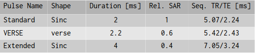
displayed in Table 1.
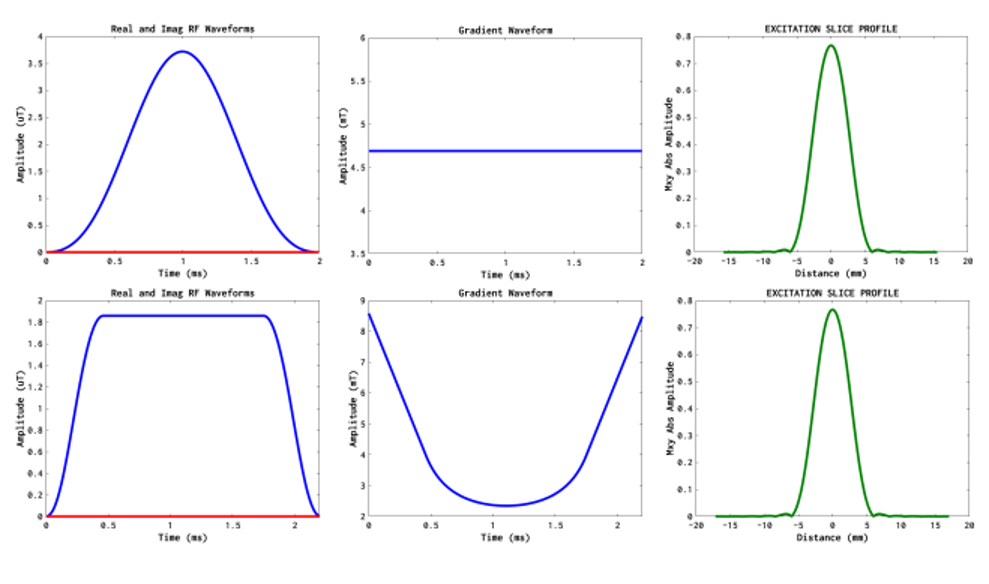
The RF pulses used in the CISS sequences are based on Hanning filtered sinc waveforms with a 1kHz bandwidth, designed in MATPULSE [6]. VERSE implementations were created using the MINVERSE algorithm [7] to reduce maximum B1 by 50%. MATPULSE was also used to simulate slice profiles, displayed in Figure 1.
Results
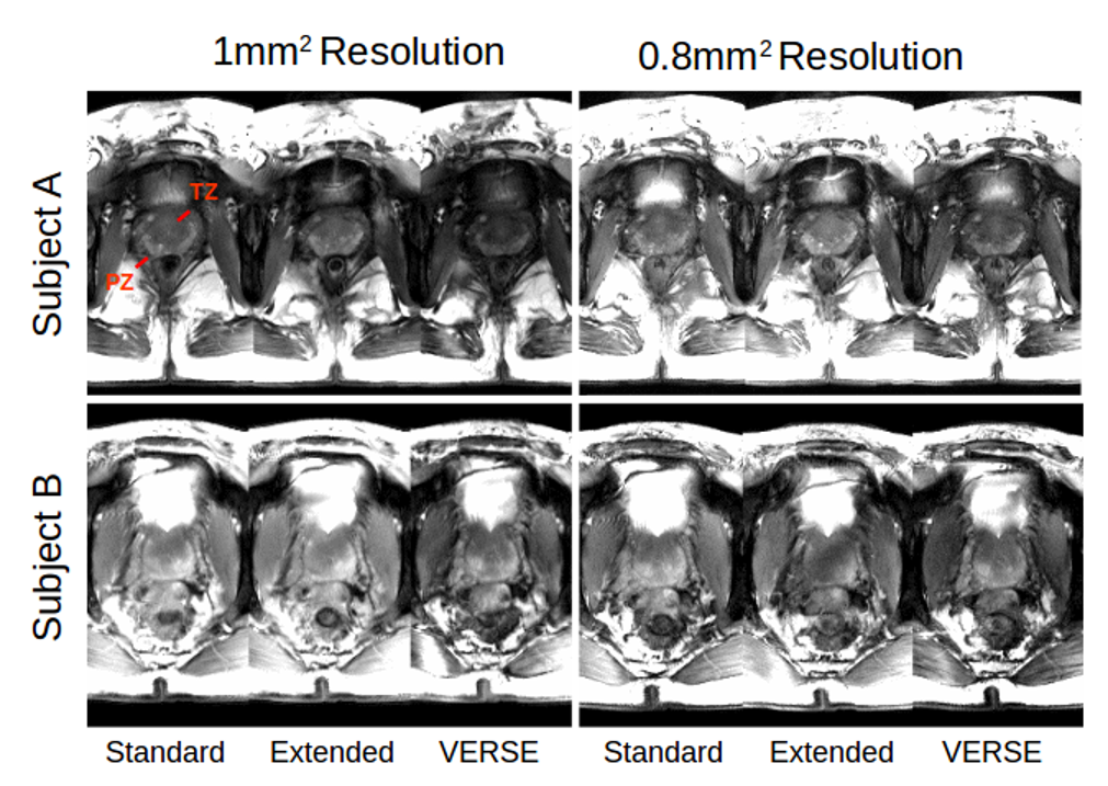
Resultant CISS images from two subjects are shown in figure 2. For the standard implementation, good contrast between the transitional (TZ) and peripheral (PZ) zones is achieved. In the extended RF implementation, contrast is decreased and in some cases, weak banding artifacts are apparent. In the VERSE implementation, SNR is visibly reduced, but contrast remains between the TZ and PZ. High signal intensity and artifacts originating from the bladder were found in some cases to hinder prostate visualization.
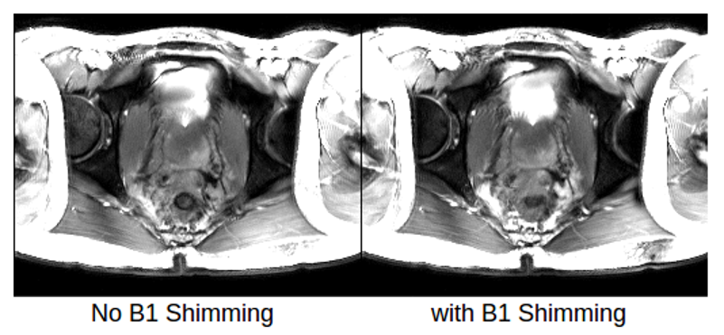
An example of the effects of local RF shimming are shown in figure 3, in which contrast is gained between the TZ and PZ after shimming, due to a local increase in the flip angle in the prostate.
Discussions
In this study, the feasibility of SSFP imaging of the prostate at UHF was investigated. SSFP, in combination with RF shimming was shown to provide image quality and contrast comparable to that reported for TSE imaging [1,2]. The use of VERSE pulses allows for a low-SAR implementation of the CISS sequence, while maintaining a short TR and minimizing off-resonance effects. Increasing the RF pulse duration increases the overall TR and the risk of artifacts due to banding.
Phase-only RF shimming was used iso as not influence the magnitude of the VERSE pulses. However, varying the amplitude in addition to phase was not found to enhance the efficiency of the RF shim.
SSFP produces a bright water signal that influences the dynamic range of the image, and signal from the bladder was found to hinder image contrast. As such, appropriate subject preparation is required before scanning.
The RF pulse design in this study was based on the default pulses of the CISS sequence. Increasing the bandwidth-time product would allow for better definition of the slice profiles, which in combination with VERSE pulses allows for the possibility of multi-slice or 3D imaging, the application of which is the subject of further investigation.
Conclusion
Prostate imaging at UHF using SSFP with RF shimming is an alternative approach to TSE approaches. SSFP produces images of comparable quality and contrast compared to those acquired with TSE. The use of VERSE pulses in this study reduces SAR by approximately 40% which would allow for greater coverage or spatial resolution.Acknowledgements
No acknowledgement found.References
[1] Metzger et al. MRM 59(2); 2008
[2] Maas et al. MRM 71(5); 2014
[3] Casselman et al. Am. J. Neuroradiol 14; 1993
[4] Orzada et al., Proc Intl. Soc MRM #2999; 2009
[5] Van de Moortele and Ugurbil, Proc Intl. Soc MRM #367; 2009
[6] Matson, MRI 12(8); 1994
[7] Hargreaves et al., MRM 52(3); 2004
Figures