2561
Multi-parametric MRI evaluation of prostate cancer volume: correlation with whole mount pathology1Department of Radiology, University of Chicago, Chicago, IL, United States, 2Department of Radiology, Guangzhou Medical University, Guangzhou, China, 3Department of Pathology, University of Chicago, Chicago, IL, United States, 4Department of Urology, University of Chicago, Chicago, IL, United States
Synopsis
This study compared prostate cancer volume determined on different multi-parametric MRI sequences: T2W imaging, ADC map and DCE-MRI with whole mount pathology in 17 patients. Tumor volumes were measured on T2W images, ADC maps and DCE-MRI by 2 radiologists and compared with reference standard volume measured from pathology. While lesion volume estimated using mpMRI sequences showed good correlation with pathology, T2W and ADC significantly underestimated, whereas DCE-MRI showed no significant difference. Therefore, DCE-MRI is the most effective sequence for estimating PCa volume with the highest accuracy compared to T2W-imaging and ADC maps and has similar good correlation and precision.
Introduction
Prostate cancer (PCa) is the most common non-cutaneous cancer in men. However, a lot of patients undergo aggressive treatment such as radical prostatectomy for indolent disease, with numerous side effects. Multiparametric MRI (mpMRI) is increasingly being used for PCa diagnosis, guiding biopsies [1] and deciding treatment option such as MR guided focal therapy [2]. However, MRI has been shown to underestimate tumor volume and there is a lack of consensus of which sequence better delineates PCa lesions for volume estimation. This study investigates the effectiveness of different multiparametric MRI (mpMRI) sequences for the evaluation of prostate cancer volume compared to histological volume measured on whole mount pathology.Methods
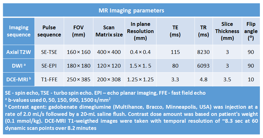
Seventeen patients (mean age = 58±7 years, PSA = 8.57±6.24 ng/ml) with biopsy confirmed PCa underwent preoperative mpMRI (see Table 1 for MR imaging protocol) with a 3T Philips Achieva MR system prior to undergoing radical prostatectomy. The prostate specimens were sectioned serially approximately in the same plane as MR images, H&E stained and evaluated by a pathologist. PCa index lesions (n=17) were marked on whole mount sections. Lesion volume was estimated using Photoshop by measuring the area on each histology slice multiplied by the slice thickness (4mm).
Two radiologists (15 and 10 years’ experience) worked in consensus to outline PCa lesions on T2-weighted (T2W) images, apparent diffusion coefficient (ADC) maps and early phase dynamic contrast enhanced (DCE) MRI. Radiologists were informed of the presence of PCa but blinded to pathology cancer outlines. The PCa lesion volumes from different mpMRI sequences were measured and compared and correlated (Pearson correlation) with reference standard volume measured from pathology. One way ANOVA with post-hoc Tukey’s test was performed to evaluate significance difference in the tumor volume estimation using histology and different mpMRI sequences and amount of underestimation between mpMRI sequences. Bland-Altman plots were performed to evaluate agreement of volume estimates using different mpMRI sequences and pathology.
Results
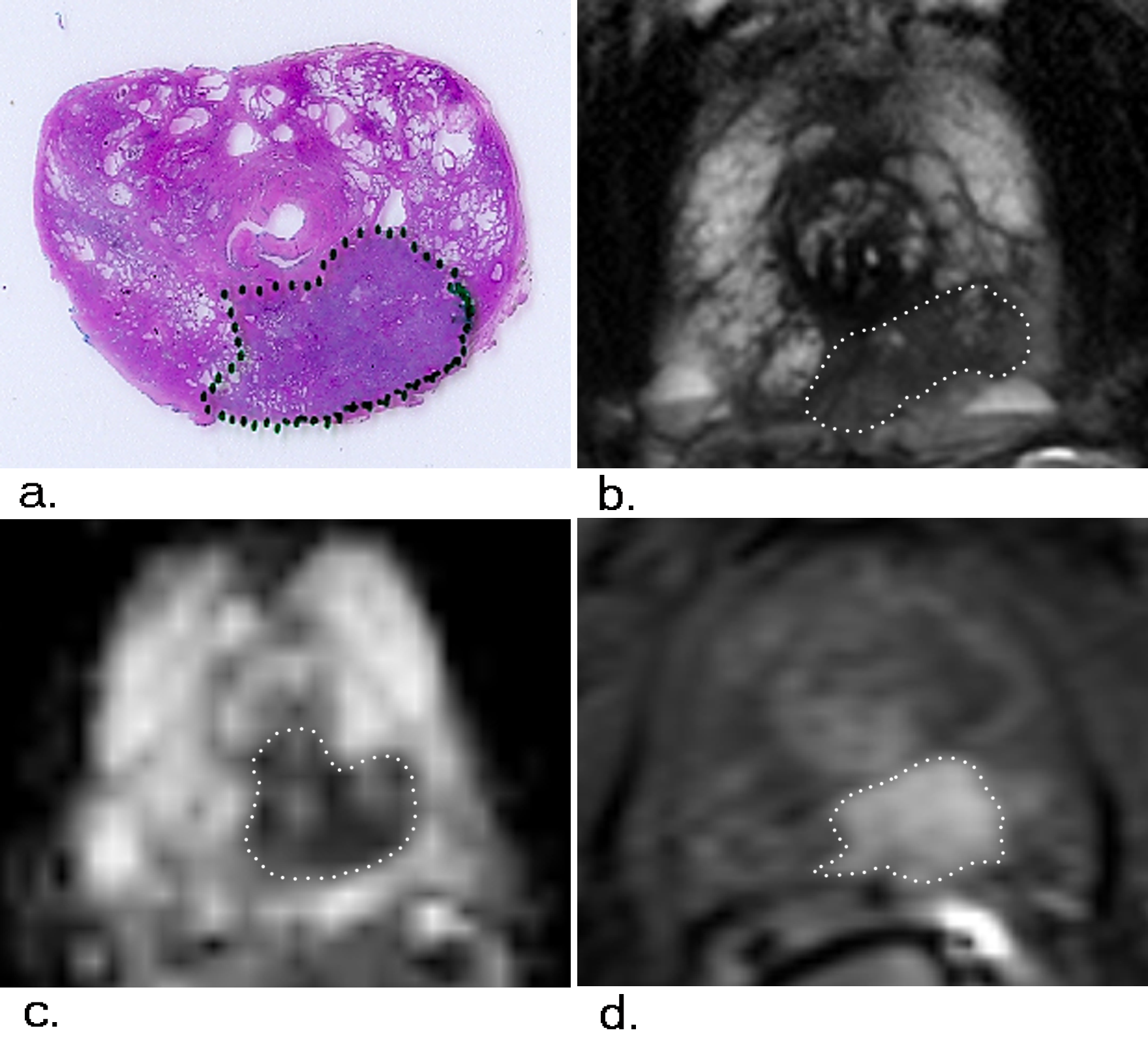
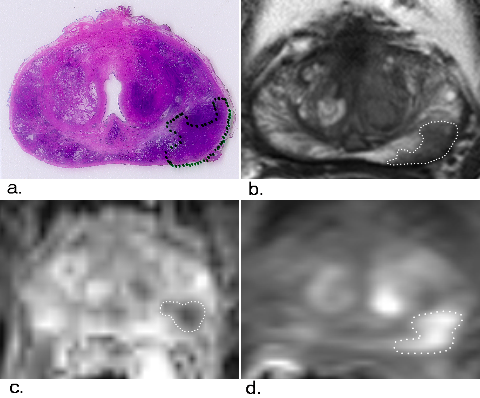
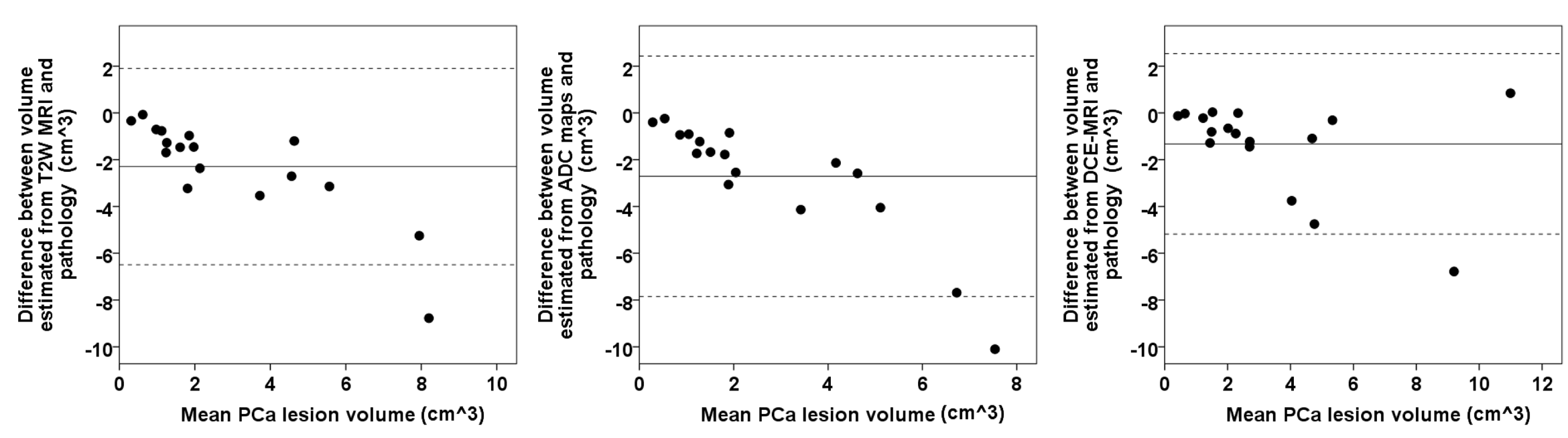
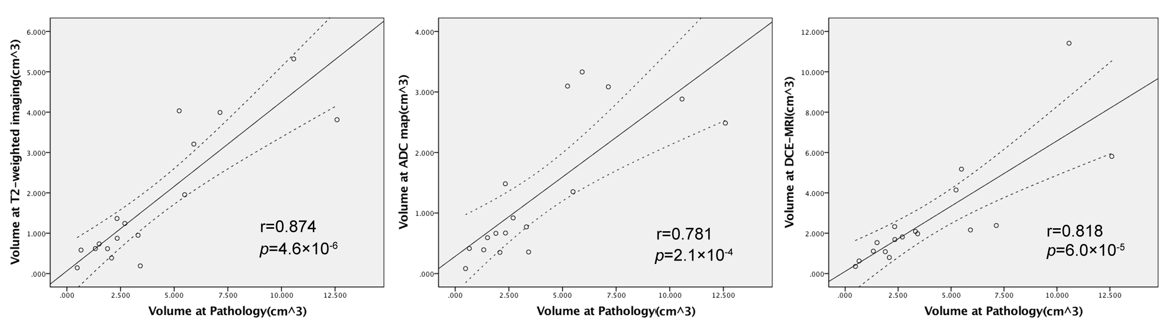
Representative examples of PCa lesions outlined on pathology and mpMRI sequences are shown in Figures 2 and 3. The mean PCa lesion volume estimated from pathology, T2W, ADC and DCE-MRI were 4.06±3.42, 1.77±1.60, 1.35±1.15 and 2.73±2.70 cm3 respectively. T2W (p=0.03) and ADC (p=0.01) showed significantly different lesion volume compared to reference standard volume from pathology, whereas DCE-MRI (p=0.38) showed no significant difference (see Bland-Altman plots in Figure 4). In addition, T2W (56.2±20.3%) and ADC (65.2±16.9%) underestimated PCa volume significantly (p<0.05) more than DCE-MRI (29.1±24%). The correlations of lesion volume estimate from T2W, ADC and DCE-MRI with pathology were 0.874, 0.781, and 0.818 (p<0.05) respectively (Figure 5).Discussion
The results suggest that DCE-MRI has good accuracy for estimation the PCa volume, whereas T2W-imaging and ADC significantly underestimate PCa volumes. While most studies [3], [4] report that MRI underestimates PCa volume, the results from our studies using DCE-MRI match that of a recent study that reported only volume was underestimated in only 8% of lesions [5]. Some studies including the previous study did not report the mpMRI sequence that they used to estimate PCa volume, or have reported volume estimated using DCE-MRI [3].
In our study, DCE-MRI shows similar good correlation in estimating volume as T2W and ADC. However this results is different from another study [4] where DCE-MRI was shown to have a bad correlation with histology for estimating PCa volume.
In addition, the DCE-MRI has similar precision as other mpMRI sequences in estimating PCa volume as evidenced by the similar standard deviation for percentage difference in tumor volume estimated from histology.
Conclusion
DCE-MRI is the most effective sequence for estimating PCa volume with the highest accuracy compared to T2W-imaging and ADC maps and has similar good correlation and precision.Acknowledgements
The study was funded by Philips Healthcare and National Institutes of Health (NIH R01 CA172801 and NIH 1S10OD018448-01).References
1. Schoots, I.G., M.J. Roobol, D. Nieboer, et al., Magnetic resonance imaging-targeted biopsy may enhance the diagnostic accuracy of significant prostate cancer detection compared to standard transrectal ultrasound-guided biopsy: a systematic review and meta-analysis. Eur Urol, 2015. 68(3): p. 438-50.
2. Mathew, M.S. and A. Oto, MRI-guided focal therapy of prostate cancer. Future Oncol, 2017. 13(6): p. 537-549.
3. Le Nobin, J., C. Orczyk, F.M. Deng, et al., Prostate tumour volumes: evaluation of the agreement between magnetic resonance imaging and histology using novel co-registration software. BJU Int, 2014. 114(6b): p. E105-e112.
4. Isebaert, S., L. Van den Bergh, K. Haustermans, et al., Multiparametric MRI for prostate cancer localization in correlation to whole-mount histopathology. J Magn Reson Imaging, 2013. 37(6): p. 1392-401.
5. Borofsky, S., A.K. George, S. Gaur, et al., What Are We Missing? False-Negative Cancers at Multiparametric MR Imaging of the Prostate. Radiology, 2017: p. 152877.
Figures