2546
Respiratory binning showdown; self-gated, respiration belt or pencil beam?1Radiology and Nuclear Medicine, Academic Medical Center Amsterdam, Amsterdam, Netherlands, 2Biomedical Engineering & Physics, Academic Medical Center Amsterdam, Amsterdam, Netherlands
Synopsis
In body MR many acquisitions respiratory motion correction is of great importance. In this study we compared self-gated motion-state binning to binning by both the pencil-beam navigator and a respiration belt by looking at the resulting image quality for each method. A 3D T1-weighted radial stack-of-stars turbo field echo (TFE) was acquired in three volunteers. The self-gated respiratory motion binning outperformed the other two methods in image quality and smoothness between the respiratory states. More subjects should be included in the study but for now it can be concluded that self-gating would be the preferred method of respiratory binning.
Purpose
In body MR many acquisitions are performed during breath-hold to minimise motion artefacts. However, this limits the acquisition time to the total time the subject can hold their breath: hence acquisition resolution is often sacrificed to decrease scantime. Alternatively, a multi-slice acquisition can make use of multiple breath-holds to increase scan resolution.1 However, motion artefacts can still be present since the volume of air in the lungs can differ between breath-holds.
To overcome this issue, gating methods can be used, ordering the data into several motion-states according to the respiratory motion, thereby reducing motion artefacts. There are several techniques to measure this respiratory signal, including a respiration belt and a pencil beam navigator.2,3 Another method to determine the respiratory motion was introduced by applying a radial stack of stars acquisition and using the centre of k-space to reconstruct the respiratory signal.4–6 This technique, called self-gating, has the advantage that no additional acquisition (pencil-beam navigator) or device (respiration belt) is needed to acquire the respiratory signal. However, little research has been performed on comparing self-gated respiratory binning to already existing methods of acquiring the respiratory motion.
In this study the self-gated motion-state binning is compared to binning by both the pencil-beam navigator and respiration belt by looking at the image quality for each method.
Materials and Methods
Subjects: The liver of three healthy volunteers were scanned (one female and two male, age=26±2.3 years) on a 3T Ingenia whole-body MR scanner (Philips Healthcare, Best, Netherlands) using a 16 channel anterior and 12 channel posterior coil. One of the volunteers was instructed to hold their breath in mid acquisition and start breathing irregular in the last segment of the acquisition to study the robustness of the methods against irregular breathing.
MR Acquisition: A 3D T1 weighted radial stack-of-stars turbo field echo (TFE) was acquired with the imaging parameters in figure 1. Raw data was exported and pre-processed using the MRecon software package (GyroTools, Zurich, Switzerland)
Self-gating respiratory signal extraction: The self-gating respiratory signal was extracted using principal component analysis on the centre of k-space.7 An iFFT was performed over all spokes in slice encoding direction resulting in a 2D radial k-space for every slice. A projection profile along the slice encoding direction was calculated for each radial spoke angle. The projections for all coils were then concatenated and a principal component analysis was performed on the projection. The principal component was selected in the expected respiratory frequency window, of 0.1-0.5 Hz, with the highest energy in k-space (figure 2) and set as the respiratory motion.
Pencil beam navigator and respiration belt signal extraction: Before each TFE train a pencil beam navigator shot was acquired. The respiration Phase Encoded Artefact Reduction (PEAR) belt was placed on the stomach of the subject at the height of the navel. Both the navigator and respiration belt signal were down-sampled to the sampling rate of the self-gating signal.
Binning and compressed sensing reconstruction: The respiratory data were binned in five equally sized motion states according to each of the three respiratory signals. The spokes were divided according to their relative amplitude in respect to the whole respiratory signal. The under-sampled data was reconstructed with a compressed sensing method using a total variation minimization along the respiratory binning dimension as described by Feng et al.7
Data analysis: The resulting images were scored by a radiology resident (7 years of MRI liver experience) looking at overall image quality and smoothness of the different respiratory states using the five point Likert scale (1-very bad, 5-very good).
Results
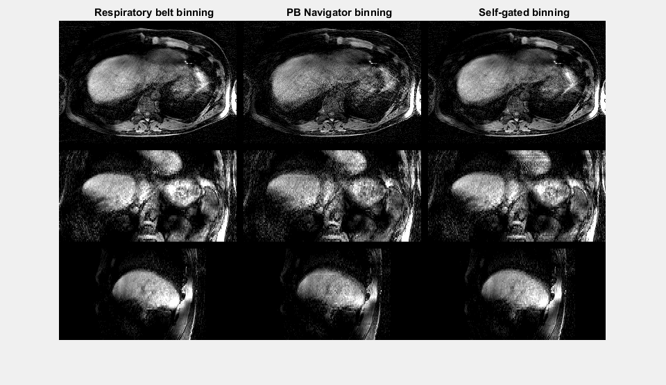
In figure 3 the respiratory signals are shown for a single volunteer were the different colours represent the five respiratory bins. To the right the corresponding reconstructed image is shown for the end exhalation bin. Looking at the smoothness of the respiration images the self-gated respiration binning shows the best results with 4 points for all volunteers. Also when looking at image quality the self-gating method outperforms the two other methods. In figure 5 a movie is shown through the respiratory motion states for the three methods.Discussion and Conclusion
In this study we showed that the self-gated respiratory motion binning outperforms the other two methods in image quality and smoothness between the respiratory states. One possible reason why the respiration belt performed less was that the respiration belt uses a dynamic rescaling of the signal (figure 3). The lower image quality of the navigator could be explained by interference of the navigator measurement with the acquisition. Future work should include more subjects, allowing statistical testing. For now it can be concluded that self-gating would be the preferred method of respiratory binning.Acknowledgements
No acknowledgement found.References
1. Paling, M. R. & Brookeman, J. R. Respiration artifacts in MR imaging: reduction by breath holding. J. Comput. Assist. Tomogr. 10, 1080–2
2. Wang, Y., Rossman, P. J., Grimm, R. C., Riederer, S. J. & Ehman, R. L. Navigator-echo-based real-time respiratory gating and triggering for reduction of respiration effects in three-dimensional coronary MR angiography. Radiology 198, 55–60 (1996).
3. Ehman, R. L., McNamara, M. T., Pallack, M., Hricak, H. & Higgins, C. B. Magnetic resonance imaging with respiratory gating: techniques and advantages. AJR. Am. J. Roentgenol. 143, 1175–82 (1984).
4. Liu, J. et al. Respiratory and cardiac self-gated free-breathing cardiac CINE imaging with multiecho 3D hybrid radial SSFP acquisition. Magn. Reson. Med. 63, 1230–7 (2010).
5. Chandarana, H. et al. Free-breathing radial 3D fat-suppressed T1-weighted gradient echo sequence: a viable alternative for contrast-enhanced liver imaging in patients unable to suspend respiration. Invest. Radiol. 46, 648–53 (2011).
6. Feng, L. et al. Golden-angle radial sparse parallel MRI: Combination of compressed sensing, parallel imaging, and golden-angle radial sampling for fast and flexible dynamic volumetric MRI. Magn. Reson. Med. 72, 707–717 (2014).
7. Feng, L. et al. XD-GRASP: Golden-angle radial MRI with reconstruction of xtra motion-state dimensions using compressed sensing. Magn. Reson. Med. 75, 775–788 (2016).
Figures