2539
Ancillary imaging features for differentiation of hypervascular hepatic tumors on Gadoxetic acid-enhanced MR imaging1radiology, Chung-ang university hospital, Seoul, Republic of Korea, 2radiology, Samsung medical center, Seoul, Republic of Korea
Synopsis
There are many types of hypervascular tumors that need to be differentiated from hepatocellular carcinoma (HCC) including focal nodular hyperplasia (FNH), hepatocellular adenoma (HCA), neuroendocrine tumor (NET), and intrahepatic cholangiocarcinoma (ICC). Since each tumor requires different treatment strategies, awareness and recognition of reliable imaging features that help precisely distinguish among these hypervascular tumors. Since these hypervascular tumors occasionally manifest overlapping imaging features, the accurate diagnosis of these tumors can still be challenging on MRI. Therefore, we conducted this study to determine ancillary imaging features that help differentiation of hypervascular hepatic tumors on gadoxetic acid-enhanced MRI.
Introduction
Hypervascular hepatic tumors usually meet MR imaging and there are many types of hypervascular tumors that need to be differentiated from hepatocellular carcinoma (HCC) including focal nodular hyperplasia (FNH), hepatocellular adenoma (HCA), neuroendocrine tumor (NET), and intrahepatic cholangiocarcinoma (ICC). Since each tumor requires different treatment strategies, awareness and recognition of reliable imaging features that help precisely distinguish among these hypervascular tumors. Advances in MR imaging technology and development of tissue-specific contrast material such as gadoxetic acid made it possible to differentiate HCCs from other hypervascular tumors based on their typical signal intensities and enhancement pattern. Since these hypervascular tumors occasionally manifest overlapping imaging features, the accurate diagnosis of these tumors can still be challenging on MRI. Therefore, we conducted this study to determine ancillary imaging features that help differentiation of hypervascular hepatic tumors on gadoxetic acid-enhanced MRI.Methods
The study was conducted by a retrospective case-control study at a single tertiary center. One hundred forty-seven patients with pathologically proven hypervascular hepatic tumors who had undergone gadoxetic acid-enhanced MRI were enrolled in this study. Included tumors were 23 focal nodular hyperplasias (FNHs), 11 hepatocellular adenomas (HCAs), 15 neuroendocrine tumors (NETs), 25 intrahepatic cholangiocarcinomas (ICCs), and 73 hepatocellular carcinomas (HCCs). MR Imaging variables including morphologic features, signal intensity, and enhancement pattern of the tumors were analyzed for determine the differential MR imaging features. We also evaluated the diagnostic performance for accurate diagnosis of each tumor by two observers.Results
Multivariate analysis revealed that hyperintensity on ADC map (63.6%) in HCA group (P = 0.002), central hypointensity on arterial phase (AP) (93.3%) in NET group (P = 0.0001), target sign on hepatobiliary phase images (HBPI) (84%) in ICC group (P < 0.0001), presence of septum (79.5%) and capsule (83.6% ) in HCC group (P < 0.0001, P = 0.0001, respectively) were significant and independent variables for differentiating each hypervascular tumor groups over other tumor groups except FNH group. Although considerable number of FNH (85%) revealed reverse target sign (central hypointensity with ring enhancement) on hepatobiliary phase images, there was no statistical difference in multivariate analysis. Diagnostic accuracies for both observers were 98 –98.6% in FNH group, 96.6 – 98% in HCA group, 97.3 – 98.6% in NET group, 90.5 – 94.6% in ICC group, and 85.7 – 93.2% in HCC group. The κ value between two observers was 0.832 for diagnosis of hypervascular tumor groups, indicating excellent interobserver agreement.Discussion
Hypervascular hepatic tumors are commonly identified in MR imaging. Since these tumors occasionally manifest overlapping imaging features, the accurate diagnosis can be challenging on conventional MRI. Our study demonstrated that hyperintensity on ADC map in HCAs, central hypointensity on AP in NETs, target sign on HBPI in ICCs, and presence of capsule and intratumoral septum in HCCs were independent and significant ancillary features for differentiation of these hypervascular tumors on gadoxtic acid-enhanced MRI.
In our study, considerable number (82.6%, 19/23) of atypical FNHs revealed reverse target appearance on HBP compared with only one case of ICC in other hypervascular tumors. The reason of these atypical findings on HBP in FNH is presumably associated with presence of central scar and absence of OATP8 expression in the hepatocytes surrounding the central scar in hypointense area, and strong expression of OATP8 in peripheral areas. Although this imaging feature were not demonstrated as significant variable, we believe it may be beneficial to differentiate atypical FNH from other hypervascular tumors.
Conclusion
Ancillary imaging features of hyperintensity on ADC map in HCAs, central hypointensity on AP in NETs, target sign on HBPI in ICCs, and presence of septum and capsule in HCCs can be helpful for differentiation of these hypervascular tumors on gadoxtic acid-enhanced MRI.Acknowledgements
No acknowledgement found.References
1. Choi JY, Lee JM, Sirlin CB (2014) CT and MR imaging diagnosisand staging of hepatocellular carcinoma: part I. Development,growth, and spread: key pathologic and imaging aspects.Radiology 272:635–654
2. Park HJ, Kim YK, Park MJ, Lee WJ (2013) Small intrahepaticmass-forming cholangiocarcinoma: target sign on diffusionweightedimaging for differentiation from hepatocellular carcinoma.Abdom Imaging 38:793–801
3. Park HJ, Jang KM, Kang TW et al (2016) Identification of imagingpredictors discriminating different primary liver tumours in patientswith chronic liver disease on gadoxetic acid-enhanced MRI: a classificationtree analysis. Eur Radiol 26:3102–3111
4. L. Grazioli, M. P. Bondioni, H. Haradome et al. (2012) Hepatocellularadenoma and focal nodular hyperplasia: value of gadoxeticacid-enhanced MR imaging in differential diagnosis. Radiology 262:520–529
5. Jlian Arista-Nasr, Jose Antonio Fernandez-Amador (2010) Neuroendocrine metastatic tumors of the liver resembling hepatocellular carcinomas. Annals of hepatology 9:186-1916. Van der Hoef M, Crook DW, Marincek B et al. (2004) Primaryneuroendocrine carcinomas of the liver: MRI features in two cases. Abdom Imaging 4:77–81
Figures
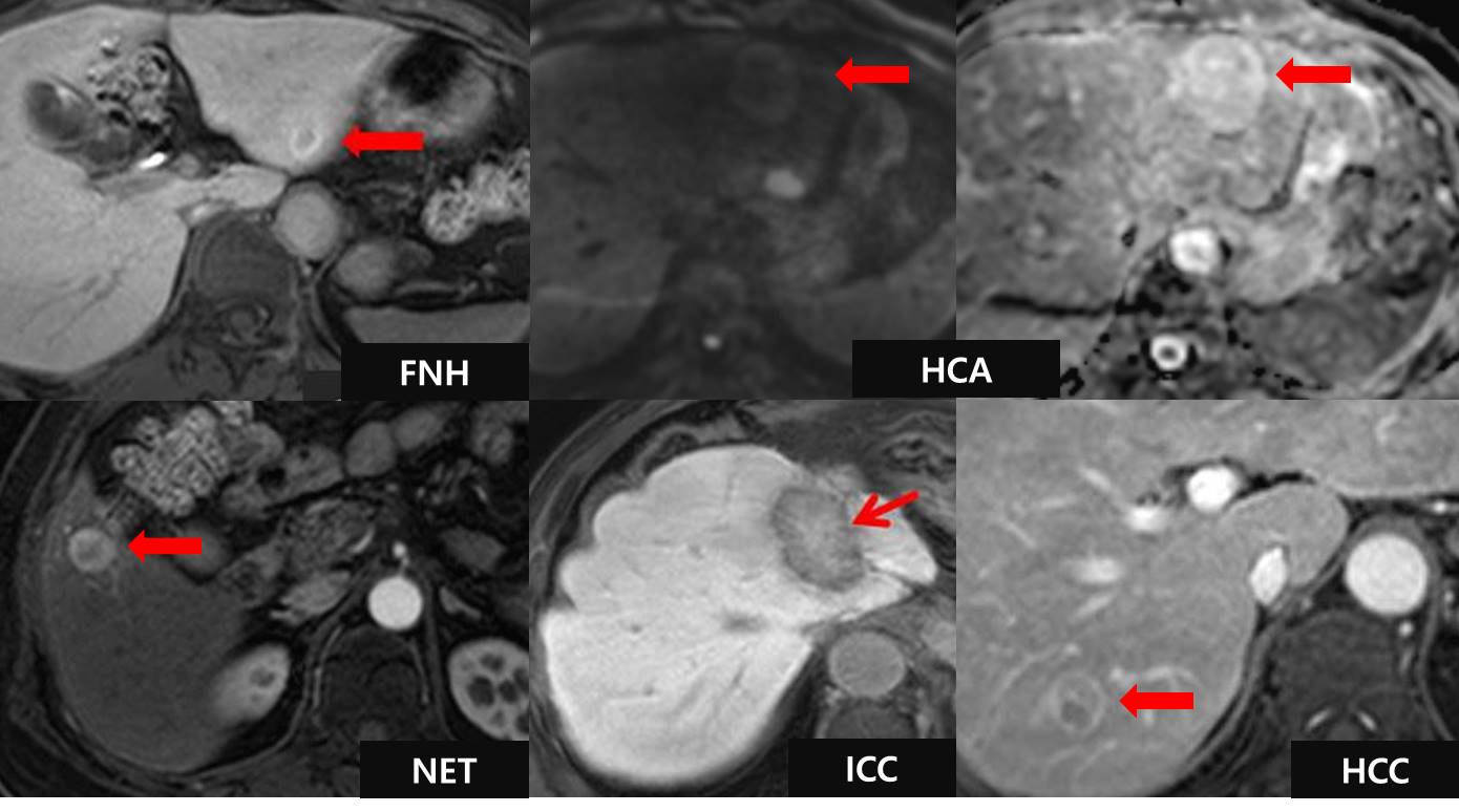
Ancillary imaging features of hypervascular hepatic tumors
Reverse target sign as central hypointensity with peripheral hyperintense rim on hepatobiliary phase image (HBPI) in focal nodular hyperplasia, hyperintensity on DWI and ADC map in hepatocellular adenoma, arterial central hypointensity in neuroendocrine tumor, target sign as central hyperintensity with peripheral hypointense rim on HBPI in intrahepatic cholangiocarcinoma, and presence of capsule and intratumoral septum in hepatocellular carcinoma are helpful imaging features to differentiate these hypervascular tumor groups.