2538
Combination of compressed sensing and two-dimensional parallel imaging can reduce the scan time for arterial phase image of gadoxetic acid enhance liver MR without degradation of image quality compared to parallel imaging alone1Radiology, Seoul National University Hospital, Seoul, Republic of Korea, 2Philips Healthcare, Seoul, Republic of Korea
Synopsis
Using combination of compressed sensing and parallel imaging for arterial phase image acquisition in gadoxetic acid enhanced liver MR
Purpose: To evaluate whether combination of compressed sensing and two-dimensional parallel imaging can reduce the scan time of triple arterial phase in gadoxetic acid-enhanced MR compared to the use of parallel imaging alone
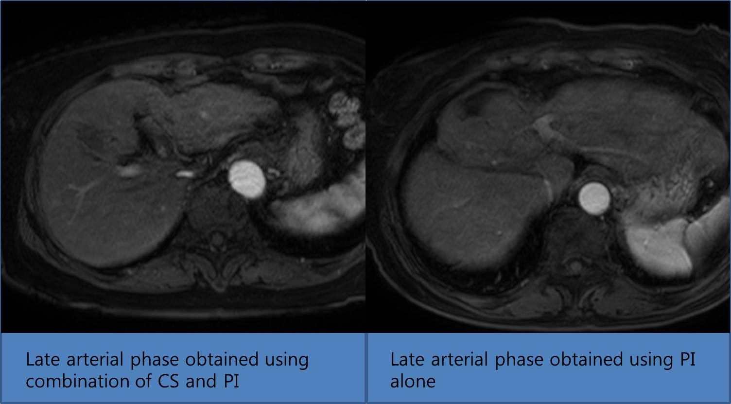
Materials and Methods: 253 consecutive patients who underwent gadoxetic acid enhanced liver MR using a 3T MR scanner (Ingenia CX; Philips Healthcare) were included in this study. Triple arterial phase (TAP) images were acquired in all patients with two different method: combination of compressed sensing (CS) and two-dimensional parallel imaging (PI) with the scan time of 15 seconds (n=127); and the use of two-dimensional PI alone with scan time of 18 seconds (n=126). Arterial phase timing was evaluated, and the degree of motion related artifact as well as image quality were assessed by using four-point scale. Scores for motion related artifact and image quality were compared between two methods using Mann-Whitney U test. The rate of successful late arterial phase acquisition without significant motion (score ≤ 2) was also calculated and compared between two methods using the chi-square test.
Results: Motion related artifact was not significantly different between two acquisition methods (3.2 for combination of CS and PI vs. 3.3 for PI alone, P=0.136). Image quality of arterial phase image obtained using combination of CS and PI (3.2) was not significantly different from PI alone (3.3) (P=0.168). TAP using combination of CS and PI captured late arterial phase without significant motion in 87.4% (111/127), and triple arterial phase using PI alone in 92.1% (116/126): this difference was not statistically significant (P=0.22).
Conclusion: Combination of CS and PI can reduce the scan time for TAP of gadoxetic acid enhanced liver MR providing comparable imaging quality to the use of PI alone.
Acknowledgements
Eun Ju Kim in Philips healthcare helped technical writing of this abstract.References
No reference found.Figures