2537
Hemodynamic Evaluations of Hepatic Vasculatures using 4D-PCA and MRFD for Liver Disease Assessment1Advanced Biomedical Imaging Research Center, Kobe University Graduate School of Medicine, Kobe, Japan, 2Center of Radiology and Radiation Oncology, Kobe University Hospital, Kobe, Japan, 3Radiology, Kobe University Graduate School of Medicine, Kobe, Japan
Synopsis
4D-PCA and MRFD can characterize liver vessels and measured WSSs provide additional information in liver disease assessments.
Background & Purpose
Liver has a unique hemodynamic system which is neither fully recognized nor understood. Shear stresses to the liver and its vessels have been reported to play important roles to keep liver function and control its regeneration. Non-contrast MRA using 4D phase-contrast angiography (4D-PCA) becomes clinically available and hemodynamic assessments using its cine data is reportedly useful for evaluations of cerebral and aortic aneurysms. In this study, we applied these techniques to hemodynamic assessments of whole liver vessels.
The purpose of this study was to evaluate the capability of four-dimensional phase-contrast angiography (4D-PCA) and magnetic resonance fluid dynamics (MRFD) for liver vessel and disease assessments
Methods & Materials
52 patients (36 men, 16 women, mean 65.9 years), who were suspected to have hepato-biliary-pancreatic malignancy and underwent 3T-MRI, were enrolled. The patients underwent MRI due to the diseases below.
- HCC:18, liver meta:10, hemangioma:1, hepatitis:2, bile duct Ca:1, PSC:1, GB stone:1, papillary Ca:1, pNET:1, panc Ca:12, IPMC:1, and IPMN:3
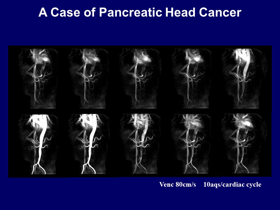
4D-PCA were obtained 3T-MR Units (Ingenia/Acheiva 3T,Philips Healthcare) (TR/TE/FA: 4.1/2.4/10, matrix: 240x191 (ZIP256x256), FOV: 400mm, thk: 120mm, slice number: 60, voxel: 1.6x1.6x2.0mm, NEX: 1, PI: 3.0 (RL), 10 aqs/cardiac cycle, VENCs of 30 and 80 cm/s, scan time: 6-10min, pulse gating).
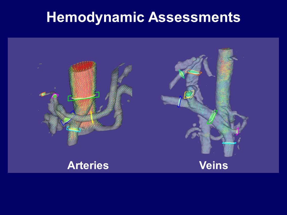
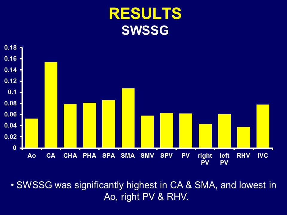
Blood flow, velocity, and shear stresses to vessel wall such as wall shear stress (WSS, Pa), oscillatory shear gradient (OSI), spatial WSS gradient (SWSSG, Pa/mm), and gradient oscillatory number (GON), were performed on MRFD software for abdominal aorta, celiac, common and proper hepatic, superior mesenteric, splenic arteries, main, right, left portal, superior mesenteric, splenic, hepatic veins, and inferior vena cava.
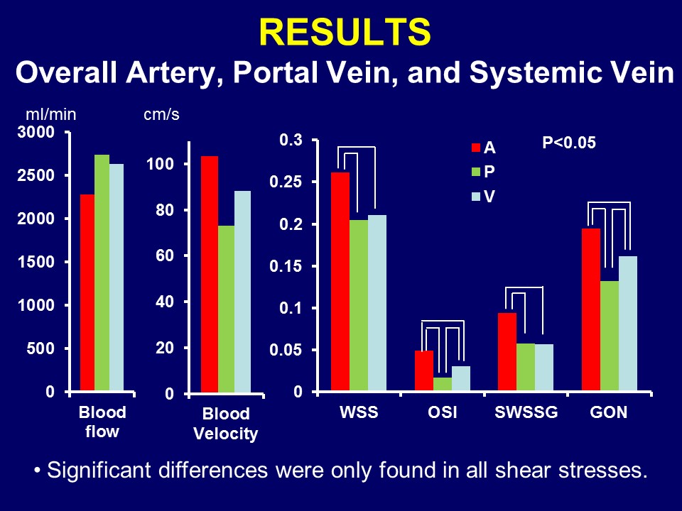
The values were compared among overall artery, portal vein, and systemic vein, and among all vessels. Correlations between the values and Child-Pugh score were assessed.
Results
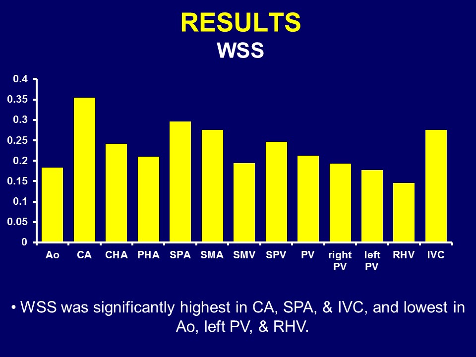
Hemodynamic assessment could be done in all vessels except for PHAs <4mm. Significant differences were only found in all shear stresses in vessel type comparisons. Blood flow and velocity were significantly highest in aorta and lowest in PHA. WSS and SWSSG were significantly highest in CA and lowest in RHV. OSI was significantly highest in aorta and lowest in lt. PV. GON was significantly highest in aorta and lowest in SMV. Significant positive correlation to CPS was found in flow, velocity, WSS, SWSSG of PHA, WSS of SPA, and flow of left PV, and negative one in GON of RHV.Discussion
4D-PCA can be clinically used as a non-contrast angiography. Further improvement of visualization is required for tiny abdominal vessels. Hemodynamic assessments for each hepatic vessel could be done in relatively short time. 4D-PCA and MRFD enables detailed hemodynamic assessment and has the potential to be used for liver disease assessments. Optimal assessment scheme is still unknown.Conclusion
4D-PCA and MRFD can characterize vessels and WSSs provide additional information in liver disease assessments.Acknowledgements
No acknowledgement found.References
- Mano Y, et al. Eur J Vasc Endovasc Surg. 2013.
- Cheng CP, et al. Am J Physiol Heart Circ Physiol. 2003.
- Stalder AF, et al. Magn Reson Med. 2008.
- Greve JM, et al. Am J Physiol Heart Circ Physiol. 2006.
- Piva A, et al. Scand J Gastroenterol. 2012.
- Wei W, et al. World J Gastroenterol. 2017.
Figures