2534
Comparison of quiet diffusion-weighted imaging with standard DWI in the abdomen: preliminary evaluation in the assessment of abdominal organs1Shandong Medical Imaging Research Institute,Shandong University, Jinan, China, 2Shandong Medical Imaging Research Institute, Jinan, China, 3Siemens Healthcare, MR Collaborations NE Asia, Beijing, China, 4Siemens Healthcare, Application Development, Erlangen, Germany
Synopsis
This study aimed to evaluate the diagnostic value of a quiet DWI (q-DWI) sequence in abdominal organs. Twenty-four patients underwent MR scans, including standard DWI and q-DWI. Quantitative and qualitative assessments regarding the signal-to-noise ratio (SNR), contrast-to-noise ratio (CNR), lesion conspicuity, the level of artifacts, and overall image quality, were measured. The qualitative rating by two radiologists shows that there were differences in lesion conspicuity, but these were not significant. The CNR and SNR of q-DWI were significantly higher than those of regular-DWI(r-DWI). For those patients who were intolerant to noise , the q-DWI technique could be more suitable.
Introduction
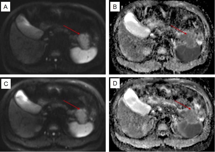
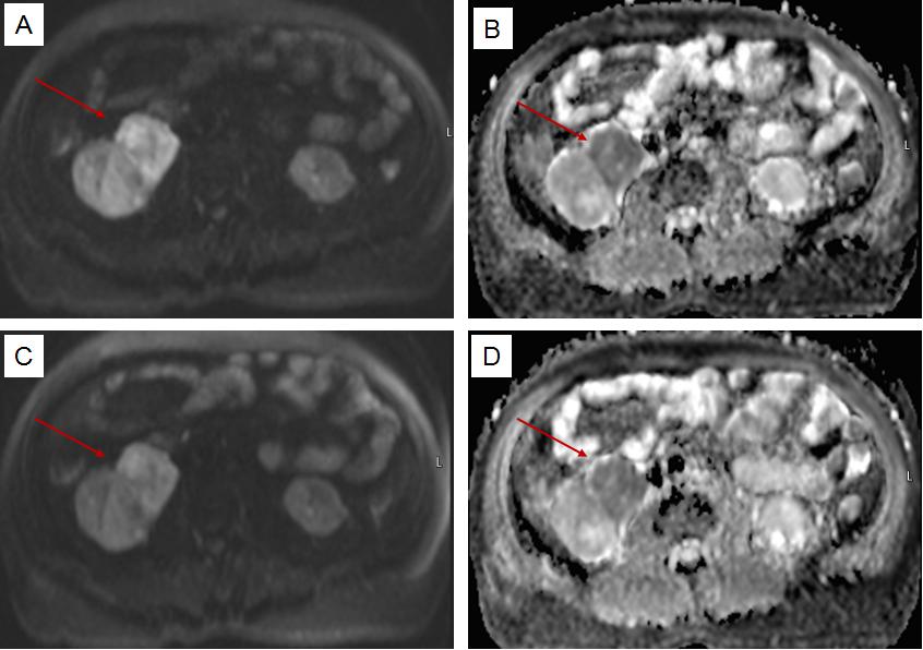
Diffusion weighted imaging(DWI) is an essential part of abdominal magnetic resonance imaging(MRI) for tumor imaging or for patients with infectious diseases, especially distinguishing enlarged lymph nodes from the surrounding tissues.1.2 However, DWI is one of the loudest MRI sequences used in clinical practice due to its high level of gradient activity, which makes patients uncomfortable and anxious, leading to more motion artifacts.3-5 Quiet DWI is a novel sequence which optimizes the gradient activity to allow for smooth and quiet gradient trajectories, e.g. by merging gradient objects or making use of available timing within the sequence. The primary purpose of our study was to evaluate and compare the quality and diagnostic value of standard single-shot EPI-based DWI (r-DWI) with that of quiet DWI (q-DWI) in MRI of the abdominal organs.Materials and Methods
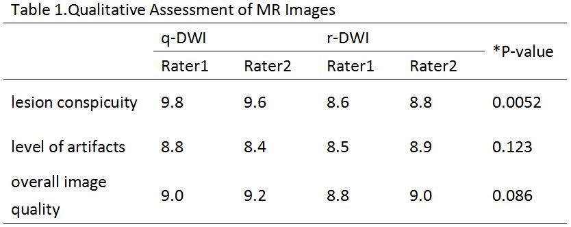
Data acquisition from twenty-four patients (45.8 ± 4.5 years old, 10 female and 14 male) with abdominal lesions was performed on a 1.5T MR system ( MAGNETOM Amira, Siemens Shenzhen Magnetic Resonance Ltd., China) with body and spine array coils. Both r-DWI and q-DWI were used to acquire the DWI images for all the patients. The two sequences had identical spatial resolution and coverage, and the scan parameters were as follows: r –DWI, TR/TE= 5000/76 ms, slice thickness = 6.0 mm, slices 25, voxel size =1.4 mm × 1.4 mm × 6.0 mm, field of view = 380 mm x 270 mm, partial-Fourier factor 6/8, bandwidth = 982 Hz/px, averages used for b = 50/b = 400/b = 800 6/9/16, acquisition time 2 min, 56 s. For q-DWI: TR/TE = 5000/84 ms, the other parameters are exactly the same. The patient datasets were rated independently by two trained radiologists blinded to the type of sequence. The qualitative evaluation included lesion conspicuity, the level of artifacts, and the overall image quality, using a grading scale score of 1 to 10, with a score of 10 indicating the most desirable quality with respect to these three criteria.6 Quantitative evaluations included an assessment of the signal-to-noise ratio(SNR) and contrast-to-noise ratio(CNR). The SNR and CNR of the different tissue types were calculated as follows:
SNRTissue1= SignalTissue1/SDNoise
and CNRTissue1Tissue2= |SignalTissue1-SignalTissue2|/SDNoise
SDNoise stands for the standard deviation in a background region.6 Inter-observer agreement for the visualization scores from the two radiologists was calculated using a kappa statistic. The scores of the two sequences in terms of the three aspects were performed by using Wilcoxon's two-sample ranked-sum test, and comparison of the categorical variables in the SNR and CNR were compared by using the independent sample T test. P < 0 .05 indicated a significant difference.
Results
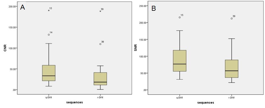
The inter-observer agreement was good both in the q-DWI (κ = 0.834) and r-DWI (κ = 0.774) images. Mean scores from the qualitative evaluation were significantly higher in q-DWI than those in r-DWI for lesion conspicuity (9.8 ± 0.6 vs. 8.6 ± 1.1, P <0 .001). No statistical difference in the level of artifacts and overall image quality was found (8.8 ± 0.5 vs. 8.6 ± 1.1; P = 0 .123; 9.0 ± 1.5 vs. 8.8 ± 0.9; P = 0.086). The detailed results regarding the qualitative analysis are shown in Table 1. The quantitative analyses revealed that the mean values of the SNR and CNR of q-DWI were higher than those of r-DWI, and the differences were significant (SNR: 90.76 ± 48.86 vs. 69.36 ± 44.68, p < 0.01; CNR: 47.73 ± 43.56 vs. 32.94 ± 40.92, p < 0.05). Figure 1 shows boxplots for the SNR and CNR values.Discussion
Our study demonstrates the feasibility of quiet MR abdominal DWI imaging at 1.5T with a comparable or higher diagnostic value compared to conventional abdominal DWI. Although the quiet sequence cannot intrinsically improve the SNR or CNR, the apparently higher SNR and CNR of the quiet sequence may be due to the high noise from the strong gradients when executing conventional DWI image acquisition that may lead to substantial motion of the patients. Considering that the number of averages of the different b-values was large, less motion will lead to a better matched average when combining the same b-value acquired at different time-points. The study findings demonstrate that the image quality of the q-DWI was comparable or superior to that of r-DWI. Lastly, a more comfortable exam environment could also benefit the abdominal MR examConclusion
The presented results may help to increase patient comfort in a routine clinical DWI examination. q-DWI is an equivalent quiet alternative to r-DWI for use in the scanning of the abdomen.Acknowledgements
No acknowledgement found.References
1. Chen ZG, Xu L, Zhang SW, Huang Y, Pan RH. Lesion discriminationwith breath-hold hepatic diffusion-weighted imaging: a meta-analysis.World J Gastroenterol 2015;21:1621–1627.
2.Choi SH, Kim KW, Lee JY, Kim KJ, Park SH. Diffusion-weighted magnetic resonance enterography for evaluating bowel inflammation in Crohn’s disease: a systematic review and meta-analysis. Inflam Bowel Dis 2016;22:669–679.
3.Foster JR, Hall DA, Summerfield AQ, Palmer AR, Bowtell RW (2000) Sound-level measurements and calculations of safe noise dosage during EPI at 3T. J Magn Reson Imaging 12:157–1634.
4.McJury M, Shellock FG (2000) Auditory noise associated with MR procedures: a review. J Magn Reson Imaging 12:37–455.
5.Quirk ME, Letendre AJ, Ciottone RA, Lingley JF (1989) Anxiety in patients undergoing MR imaging. Radiology 170:463–4666.
6.Rösch J, Ott M, Heismann B, et al. Quiet diffusion‐weighted head scanning: Initial clinical evaluation in ischemic stroke patients at 1.5 T[J]. Journal of Magnetic Resonance Imaging, 2016, 44(5): 1238-1243.
Figures
*significance level of the difference q-DWI vs. r-DWI (mean)