2532
Validation of Reproducibility of Both Zoom Diffusion Imaging And Conventional Full Field of View Method in The Kidney StudyHsuan Wen Yu1,2, Feng Mao Chiu3, Cheng Ping Chien2, and You Yin Chen1
1National Yang-Ming University, Taipei, Taiwan, 2Taipei Beitou Health Management Hospital, Taipei, Taiwan, 3Philips Healthcare, Taipei, Taiwan
Synopsis
Diffusion Tensor Imaging (DTI) is a reliable tool for investigating renal microstructure and renal function, the imaging stability remains challenging. Recently, the image-quality improvement by zoom DTI technique (reduced Field-Of-View diffusion) is reported1. We scanned 10 healthy volunteers by this technique via the respiration-triggered acquisition, and we assessed different ROIs within the medulla and the cortex of the kidney. In this study, the reproducibility between different subjects in zoom DTI was more promising when compared to full-FOV DTI. More DTI scalars were compared between zoom and full-FOV DTI in cortex and medulla and these may be potential parameters to detect pathological changes in kidney.
Introduction
Previous studies revealed that Diffusion Tensor Imaging (DTI) is a reliable tool to provide the information about renal microstructure and function1. Diffusion anisotropy in the medulla is significantly higher than the cortex, and medullary Fractional Anisotropy (FA) demonstrates a highly positive correlation with eGFR (estimated Glomerular filtration rate)1. However, only the FA and Apparent Diffusion Coefficient (ADC) values were discussed in related studies2,3, and geometric distortion and imaging blurring of diffusion tensor Echo Planar Imaging (EPI) remains challenging due to field inhomogeneity, long echo trains and the respiratory motion. The zoom diffusion imaging employs non-coplanar excitation to acquire within a small FOV, and the signal outside the FOV will not be wrapped into the image. According to recent reports, the zoom diffusion technique not only improves image spatial resolution, but also reduces the distortion artifacts2,3. The aim of this study is to assess the inter-subject reproducibility of DTI parameters, FA, Radial Diffusivity (RD), Relative Anisotropy (RA), Axial Diffusivity (AD), and ADC in zoom diffusion and full-FOV images with identical geometric parameters.Material and Methods
10 healthy volunteers (5 men and 5 women; mean age, 27±3 years) without any renal pathology were examined on a 3T MR scanner (Philips Healthcare, Eindhoven, The Netherlands), and both zoom DTI and full-FOV DTI sequences were performed in the same position. The zoom DTI uses non-coplanar excitation to acquire the signal only inside FOV, and the full-FOV DTI uses the oversampling way with additional FOVs to prevent the aliasing artifact. The following were parameters of zoom DTI and full-FOV methods respectively: TR varies with the respiratory cycle of the subjects. (Range:1-3s), TE=40/63ms, EPI factor=41/45, SENSE factor=3/1, slice thickness=6mm, FOV=140x140mm, acquisition matrix=64x64, b-values=0 and 250 s/mm2, diffusion directions=6, slice orientation=coronal, and phase encoding direction in RL. Each subject was examined with the respiratory trigger. The image analysis was performed on the workstation (IntelliSpace Portal, Eindhoven, The Netherlands). We evaluate the tensor maps (ADC, AD, RD, FA and RA maps) by measuring regions of interest (ROI) at medullary and cortical area based on b0 image. Two radiology technologists contoured all ROIs respectively, and both technologists are with 10 years of experience in radiology. The statistics analysis was executed on SPSS version 20.0 (SPSS Inc., Chicago, III)Results
Figure 1 shows a better boundary between cortex and medulla in zoom DTI. For quantitative analysis, and the averaged DTI parameters of cortex and medulla are shown in Figure 2. The averaged FA value in medulla is higher than in cortex, and the mean ADC value in medulla is lower than in cortex, and these results are similar to previous reports1,3. In addition, we observed that the ADC and RD values are higher in cortex, and the AD, FA and RA values are lower in medulla in both zoom and full-FOV DTI. It shows that the coefficient of variation (CV) of zoom DTI is lower than full-FOV method (Figure 3). The assessment of inter-observer variability were examined with paired t-tests, and there is no significant difference (all p value≥0.801).Discussion and Conclusion
We found that zoom diffusion imaging gives sharper images, even the same geometrical setting in both two imaging methods, and this might be caused by the shorter echo trains in zoom DTI. Our result showed the lower CV in zoom DTI parameters, and it could be the improvement of the image quality. It is important to get stable measurements for long-term observation, and zoom diffusion imaging could help to improve the reproducibility. This technique could be applied to early detection, treatment response and long-term observation in the renal disease.Acknowledgements
No acknowledgement found.References
- Lu, Lan, et al. "Use of diffusion tensor MRI to identify early changes in diabetic nephropathy." American journal of nephrology 34.5 (2011): 476-482.
- Notohamiprodjo, Mike, et al. "Diffusion tensor imaging (DTI) of the kidney at 3 Tesla–feasibility, protocol evaluation and comparison to 1.5 Tesla." Investigative radiology 45.5 (2010): 245-254.
- Palmucci, Stefano, et al. "Diffusion weighted imaging and diffusion tensor imaging in the evaluation of transplanted kidneys." European journal of radiology open 2 (2015): 71-80.
Figures
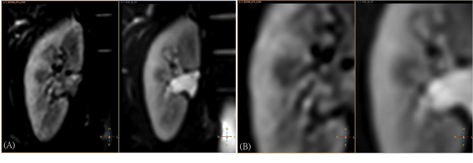
(A) DTI b=0 image performed in zoom DTI
technique. (B) DTI b=0 image performed in full-FOV methods. The boundary
between cortex and medulla was better visualized in zoom DTI.
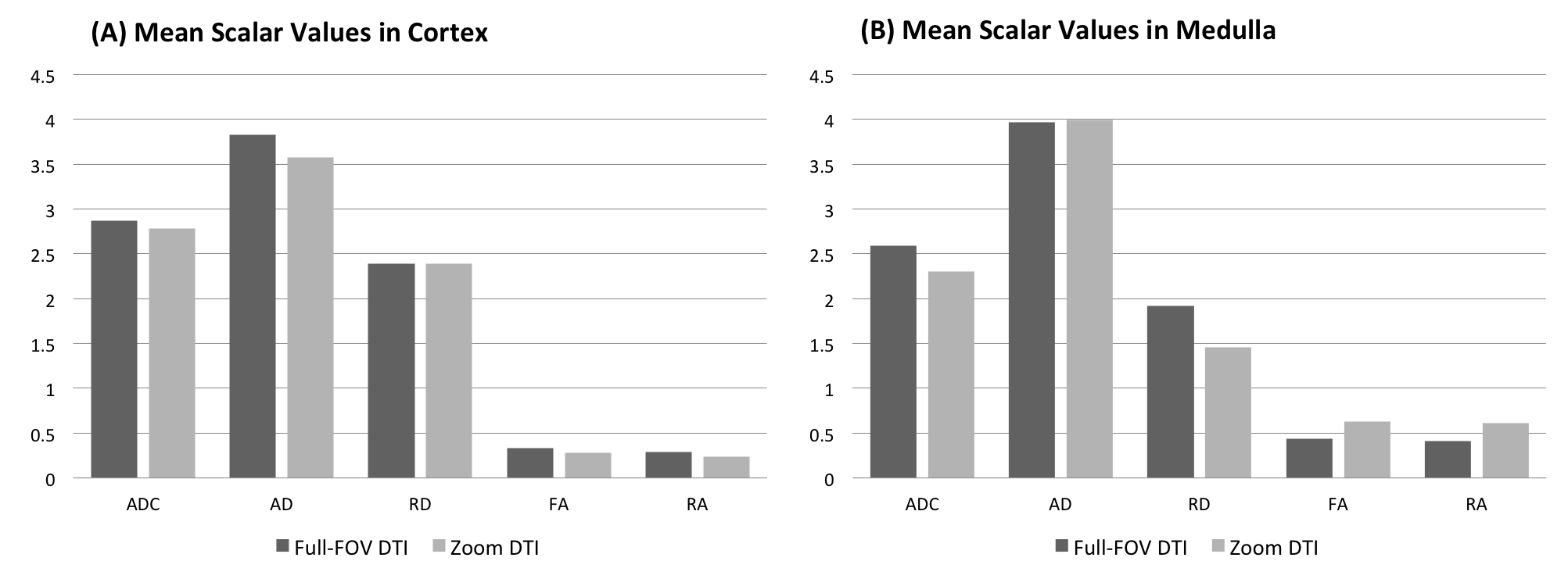
Mean scalar values in (A) cortex and (B) medulla.
ADC: (10-3 mm2/s), AD:
(10-3 mm2/s),
RD: (10-3 mm2/s).
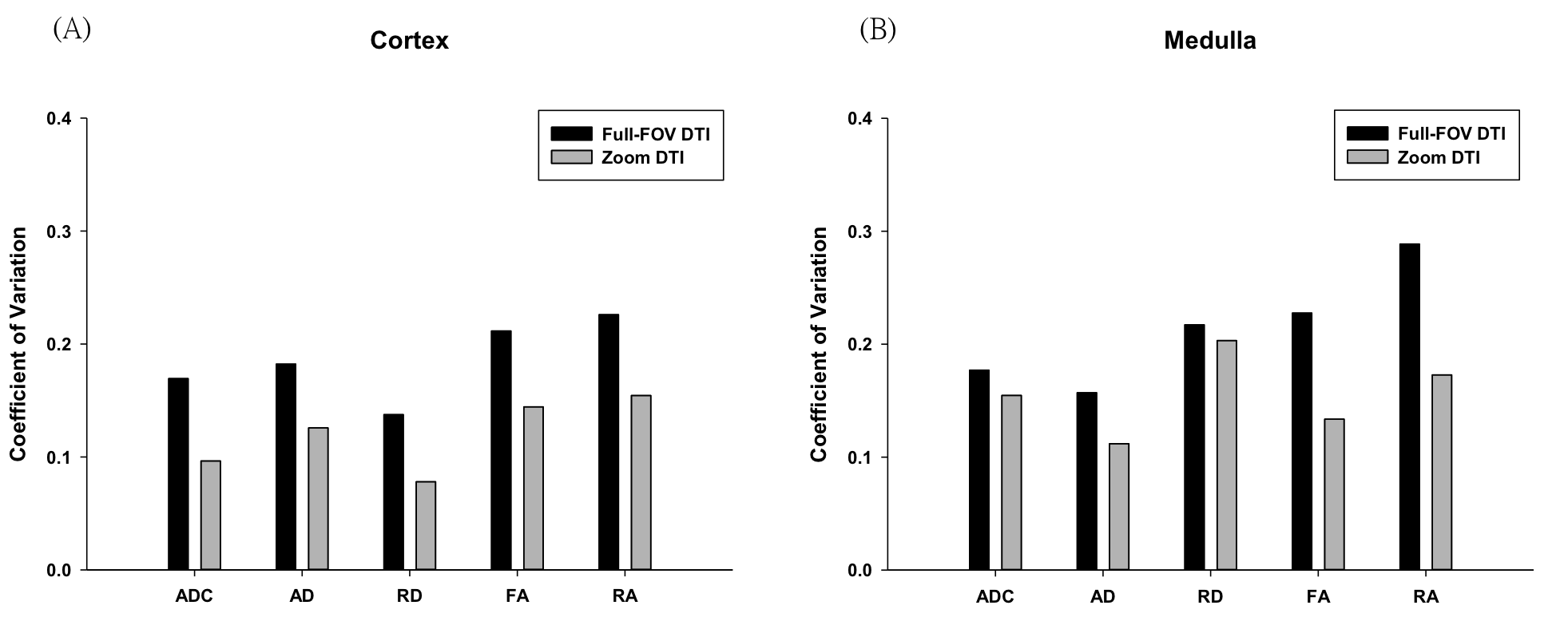
The coefficient of variance for (A) cortex and
(B) medulla, and the DTI scalars in zoom DTI demonstrates better reproducibility.