2523
Changes of T2 signal intensities of abdominal organs between pre- and post-enhanced HASTE using ferumoxytol1Radiology, David Geffen School of Medicine at UCLA, Los Angeles, CA, United States, 2Radiology, Samsung Medical Center, Seoul, Republic of Korea, 3Cardiology, David Geffen School of Medicine at UCLA, Los Angeles, CA, United States
Synopsis
This study was designed to investigate the signal intensities (SI) of abdominal organs on pre- and post-enhanced HASTE images using ferumoxytol at a dose of 4mg /kg, and to compare the differences of enhancement effect among the organs. We found that the SI of liver, spleen, and pancreas were significantly decreased on HASTE after ferumoxytol administration. The greatest effects on SI were observed in liver and spleen. Little change in SI of muscle and fat was noted. The findings suggest that normal liver and spleen undergo profound decrease in signal intensity following ferumoxytol injection, likely reflecting their high blood volume. This observation suggests a potential role for ferumoxytol in detection and characterization of focal lesions of the liver and spleen.
INTRODUCTION
Ferumoxytol is an ultrasmall superparamagnetic iron oxide (USPIO) particle, with potent T1 and T2 relaxivity and it is neither accumulated in the body nor excreted through the kidney. It is increasingly used in patients with renal impairment,1,2 with encouraging early safety results.3 Many patients with renal impairment need liver and kidney MRI, but few data address the potential use of ferumoxytol for imaging of abdominal organs. Ferumoxytol has high T1 relaxivity and the strategy for performing T1-weighted assessment of enhancement would be postulated to parallel that for the gadolinium based contrast agents (GBCA). However, ferumoxytol also shortens T2, so a strategy for performing T2-weighted enhancement imaging may be incrementally beneficial. The purpose of this study is to investigate the induced changes in signal intensity (SI) induced by ferumoxytol 4 mg/kg, on T2-weighted HASTE images of abdominal organs including liver, spleen, pancreas, muscle and fat and to determine whether this strategy is likely to increase sensitivity and specificity for focal lesions in these organs.METHODS
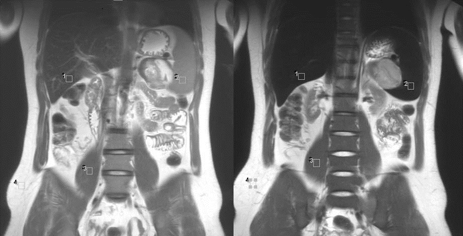
The MR images of 50 adult patients who underwent both pre- and post-ferumoxytol HASTE (mean age, 56.8 years old; M : F = 30 : 20) were analyzed. A brief scan protocol was following: MRI was performed on a 3.0-T, 32-channel, and whole-body MRI system. Pre-enhanced HASTE was obtained, and then ferumoxytol was administered intravenously in a dose of 4 mg/kg. After vascular imaging, post-enhanced HASTE was obtained. Image analysis was performed by an abdominal radiologist with 14-year experience of abdominal MRI. Quantitative analysis was performed. He drew regions of interest (ROI) on liver, spleen, pancreas, belly of psoas muscle, and subcutaneous fat layer to measure the SI of abdominal organs avoiding the large vessels and artifacts, and they were located in the same area of both images (Fig. 1). The areas of ROI were around 100 mm2. And the elongated ROI with phase encoding direction was drawn on the background to measure the standard deviation of SI which represents background noise. Given the measured values, signal-to-noise ratio (SNR) of the organs, organ-fat contrast-to-noise ratio (CNR), and the reduction in relative SNR (%SNRdecrease) were calculated.4 Paired t-tests were performed for comparison of SNR and CNR between pre- and post-HASTE images, and Kruskal-Wallis test was performed for comparison of %SNR decrease among the abdominal organs.RESULTS
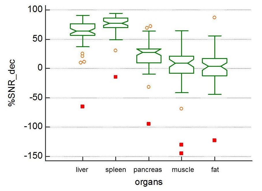
Comparing SNRs between pre- and post-HASTE, those of liver, spleen, and pancreas decreased significantly on post-HASTE image (P <0.001) (Fig. 2A). However, those of psoas muscle and subcutaneous fat were not different between two images (P = 0.316 and 0.766, respectively). Also in the analyses of organ-fat CNR, those of liver, spleen, and pancreas on post-HASTE were significantly improved (P < 0.001, < 0.001, and 0.005, respectively), but psoas-fat CNR was not improved (P = 0.480) (Fig. 2B). On the comparison of %SNRdecrease of abdominal organs, the %SNRdecrease of spleen was the lowest (74.6% ± 17.9%), which was significantly different from other organs; and followed by liver (59.8% ± 25.7%), pancreas (22.4% ± 27.4%), psoas muscle (2.2% ± 40.3%), and subcutaneous fat layer (0.6% ± 30.5%) (Fig. 3).DISCUSSION
Our results show that ferumoxytol greatly decreases the parenchymal SI of liver, spleen, and pancreas and has little effect on resting muscle and abdominal fat. These changes likely reflect the varying blood volume composition of these organs and tissues and suggest a ready mechanism for heightened T2 contrast in liver and splenic mass lesions. Therefore, ferumoxytol may enhance the detectability of focal lesions which develop in these organs, like with superparamagnetic iron oxide (SPIO).5 And this study shows the organ-fat contrast was increased, and it would be useful to depict pancreatic and gastrointestinal lesions, such as pancreatic cancer, focal pancreatitis and inflammatory bowel disease, obscured by surrounding fat or fluid more clearly. Following studies to prove the usefulness of abdominal application of ferumoxytol should be needed.CONCLUSION
The SI of liver, spleen, and pancreas were significantly decreased on HASTE after ferumoxytol administration. It had the greatest effect on the splenic SI, and followed by the hepatic, pancreas, psoas muscle and subcutaneous fat. These observations suggest a ready mechanism for heightened T2 contrast in liver and splenic mass lesions.Acknowledgements
noneReferences
1. Nayak AB, Luhar A, Hanudel M, et al. High-resolution, whole-body vascular imaging with ferumoxytol as an alternative to gadolinium agents in a pediatric chronic kidney disease cohort. Pediatr Nephrol. 2015;30(3):515-521.
2. Nguyen KL, Moriarty JM, Plotnik AN, et al. Ferumoxytol-enhanced MR angiography for vascular access mapping before transcatheter aortic valve replacement in patients with renal impairment: a step toward patient-specific care. Radiology. 2017 Oct 16. E-pub
3. Nguyen KL, Yoshida T, Han F, et al. MRI with ferumoxytol: a single center experience of safety across the age spectrum. J Magn Reson Imaging. 2017;45:804-813.
4. Yoshikawa T, Mitchell DG, Hirota S, et al. Gradient- and spin-echo T2-weighted imaging for SPIO-enhanced detection and characterization of focal liver lesions. J Magn Reson Imaging. 2006;23:712-719.
5. Toth GB, Varallyay CG, Horvath A, et al. Current and potential imaging applications of ferumoxytol for magnetic resonance imaging. Kidney Int. 2017;92:47-66.
Figures