2505
Evaluation of Simultaneous MRI/PET of Supraclavicular BAT for Detecting Adaptive Thermogenesis after Sympathetic Nervous System Activation1Laboratory of Molecular Imaging, Singapore Bioimaging Consortium, Agency for Science Technology and Research (A*STAR), Singapore, Singapore, 2Clinical Nutrition Research Centre, Agency for Science, Technology and Research (A*STAR), Singapore, Singapore, 3Singapore Institute of Clinical Sciences, Agency for Science, Technology and Research (A*STAR), Singapore, Singapore, 4Clinical Imaging Research Centre, Agency for Science, Technology and Research (A*STAR), Singapore, Singapore, 5Department of Radiology, Nationwide Children's Hospital Columbus, Columbus, OH, United States, 6Department of Endocrinology, Tan Tock Seng Hospital, Singapore, Singapore
Synopsis
There is a large interest in detecting and quantifying brown adipose tissue (BAT) in humans for evaluating its potential to design therapeutic strategies to combat obesity-related metabolic dysfunction. In the current study, we evaluated the use of simultaneous PET/MRI of
Introduction
There is a large interest in detecting and quantifying brown adipose tissue (BAT) in humans for evaluating its potential to design therapeutic strategies to combat obesity-related metabolic dysfunction[1, 2]. In contrast to white adipose tissue (WAT), which primarily store energy, BAT is thermogenic, and dissipates energy in the form of heat, on activation of the SNS[3, 4]. The gold standard technique to measure the activity of BAT is 18F-fludeoxyglucose positron emission tomography (18F-FDG PET) because of its high glucose uptake upon activation. BAT exhibits smaller multilocular adipocytes with more tissue water relative to fat than WAT, which allows the two tissues to be distinguished by Dixon-based imaging[5]. BAT fat content further decreases upon BAT activation due to fat oxidation. In the current study, we evaluated the use of simultaneous MRI/PET of the supraclavicular BAT (sBAT) in subjects with low and high adaptive thermogenesis after SNS activation by cold exposure and capsinoids ingestion[3, 4]. We also evaluated the duration of cold-exposure for changes in 18F-FDG uptake and Dixon-based fat-fraction (FF).Methods
Imaging experiments were performed on a hybrid 3T MR/PET (Biograph mMR; Siemens). MR/PET was performed simultaneously on 18 subjects with cold exposure and oral intervention of capsinoids after fasting in two separate visits. The PET scans were acquired for 80 min after 25-30 min post intravenous injection of 3 mCi (111 MBq) of 18F-FDG administered through an intravenous cannula in a vein. The MRI-FF was assessed at the end of the scan.Twelve mg of capsinoids were given before injection of 18F-FDG for the first visit of the subject. In the second visit of the same subject wore a cold thermal vest jacket (14.5 °C) for 1-2 h prior to the scan. 3-dimensional T2-weighted anatomical images was acquired using TR = 1300 ms; TE = 120 ms; FOV, 384 mm × 384 mm; matrix size 192 × 192. A 3D multi-point VIBE Dixon sequence was utilized for acquiring the water and fat images with following parameters: TR 15 ms; ten echoes time, 1.23, 2.46, 3.69, 4.92, 6.15, 7.38, 8.61, 9.84, 11.07 and 12.30 ms; FA, 4°; FOV, 384 mm × 384 mm; matrix size 192 × 192; 112 slices with 2 mm thickness. FF was obtained by fitting the multi-echo data to a 7-peak fat model using the Levenberg-Marquardt algorithm[1, 6, 7]. An affine based registration was used to register FF and PET images. Rolling ball background subtraction algorithm followed by the percentile based automatic thresholding method to exclude the unwanted region[2,8]. sBAT were manually segmented from PET and MR images based on anatomical information in multiple slice images using ITK-SNAP and under the closed guidance of expert radiologist[9]. In separate visits, energy expenditure (EE) was also measured by whole-body indirect calorimetry, at baseline and two hours after capsinoids ingestion and cold exposure. Adaptive thermogenesis was defined as the increase in EE over baseline after 2h of stimulation[10]. Subjects were stratified into high and low responders to each cold and capsinoids stimulation based on the median value of adaptive thermogenesis.Results
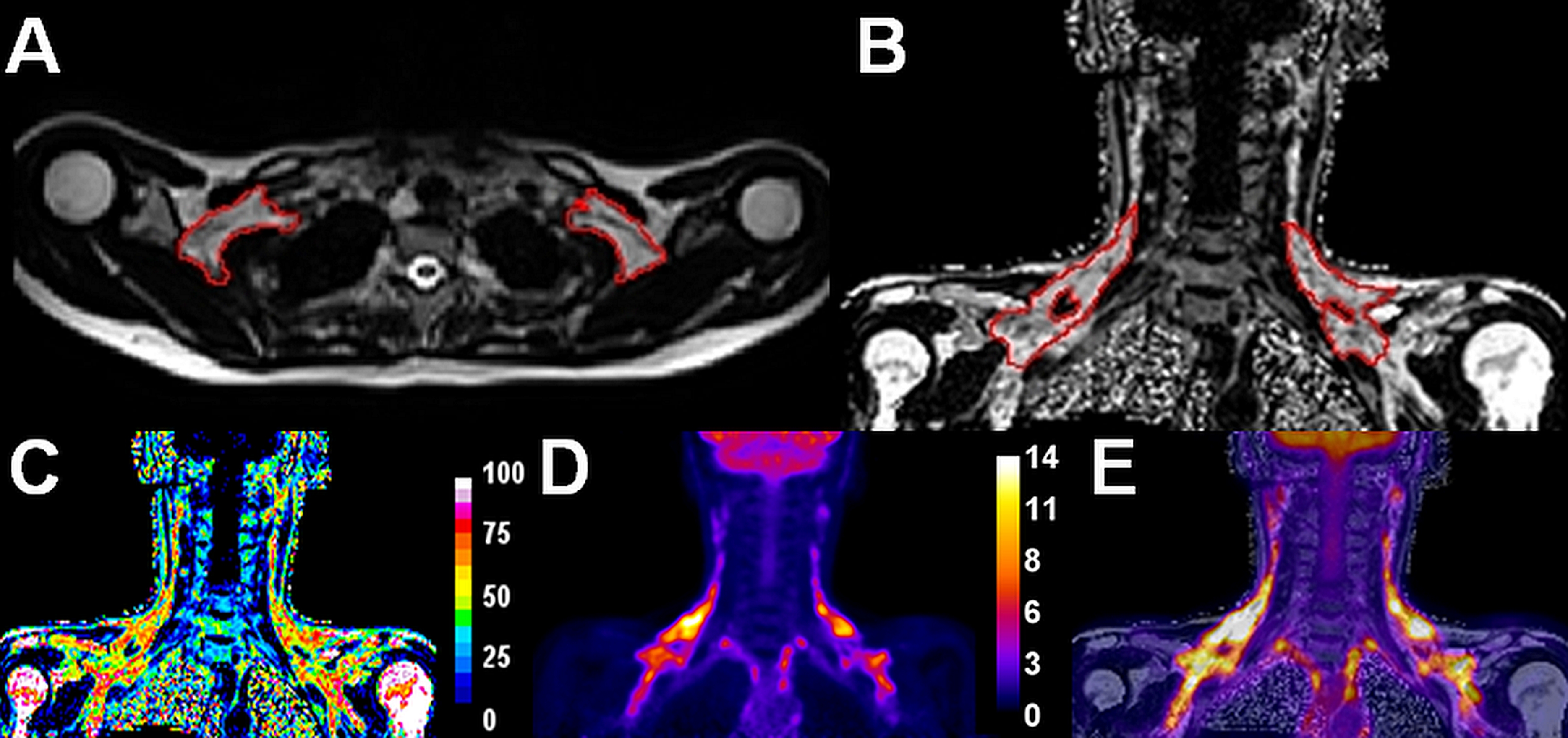
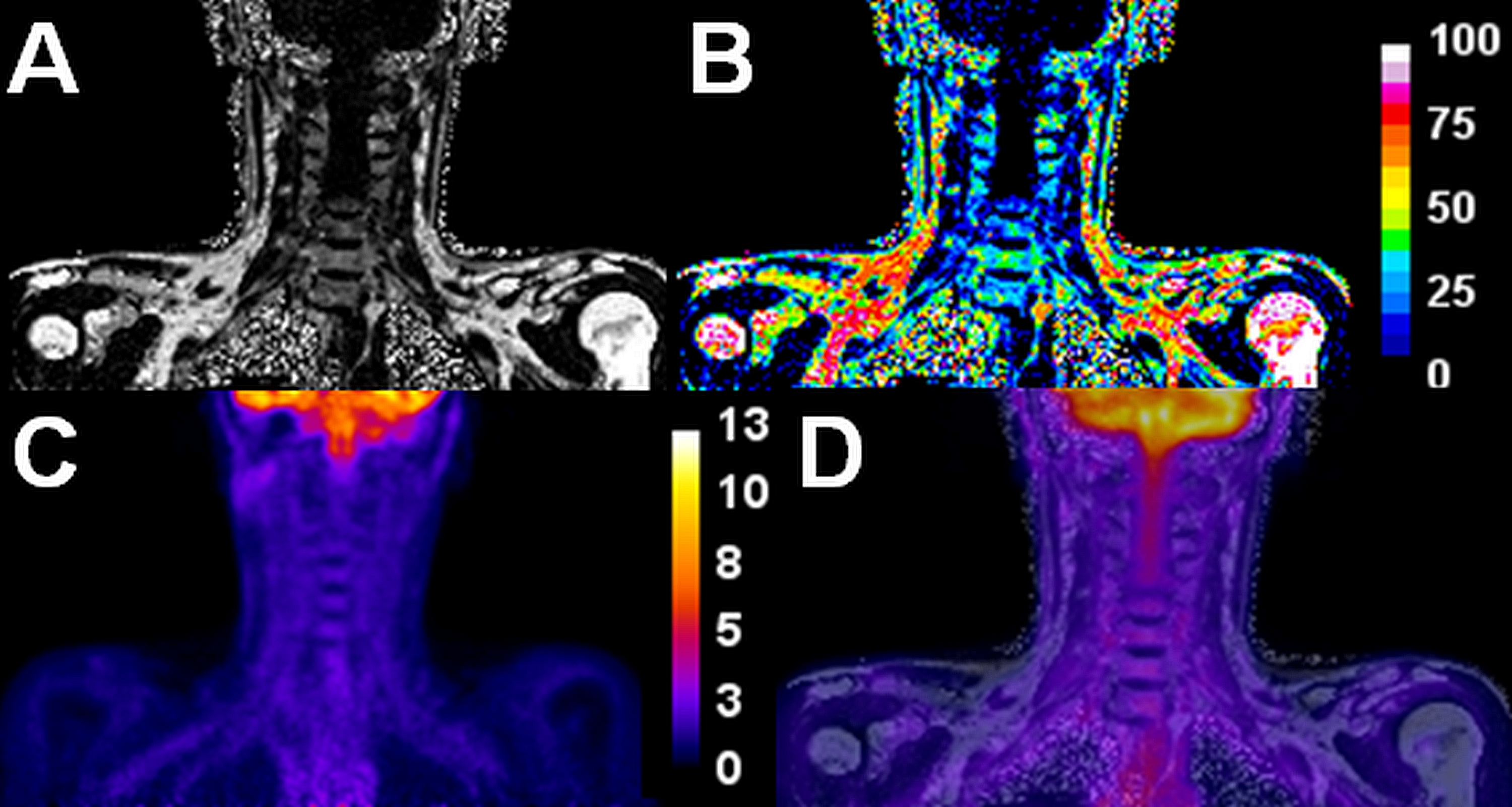
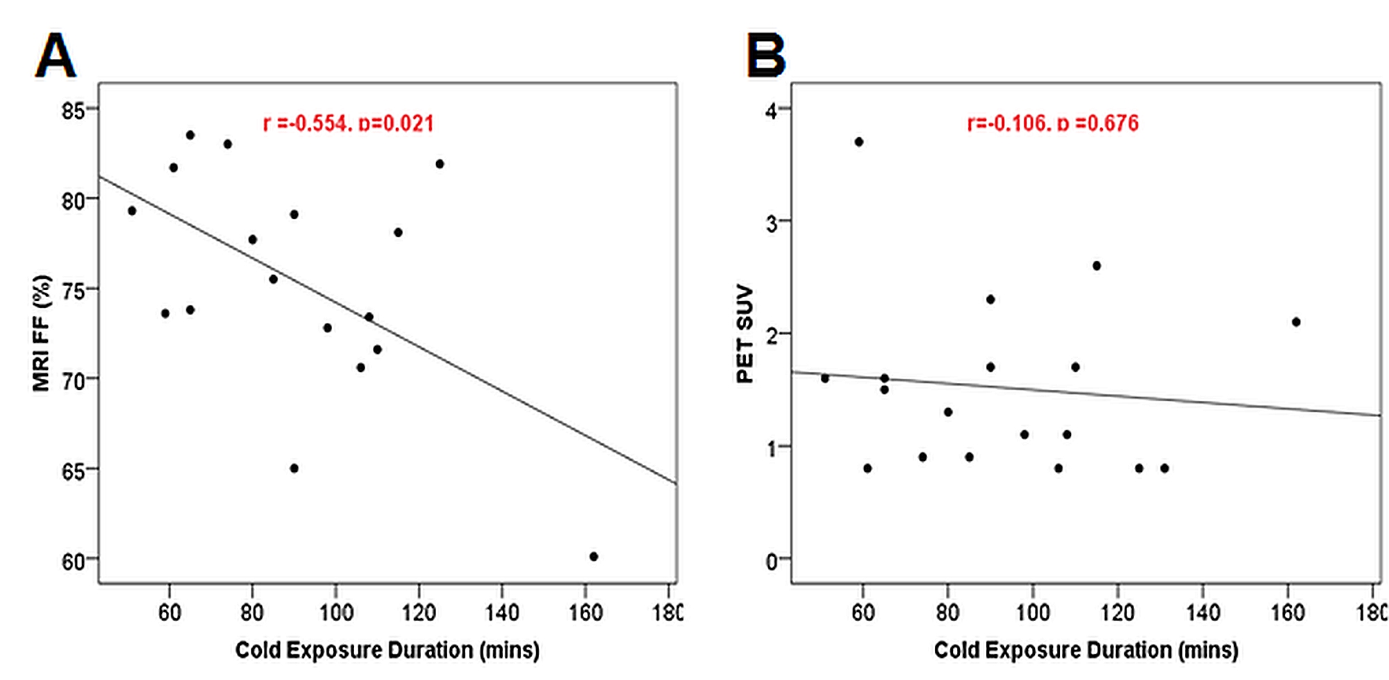
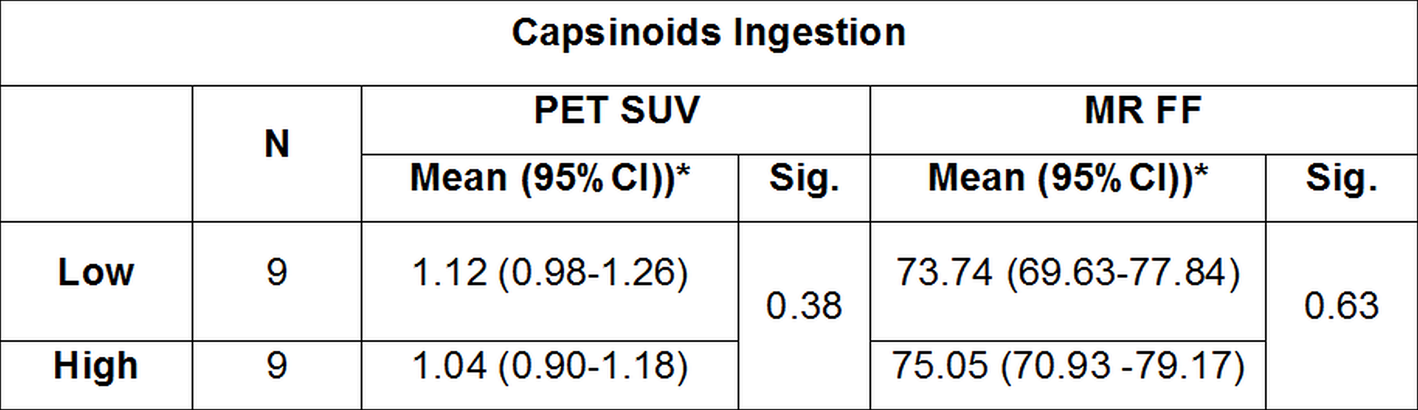
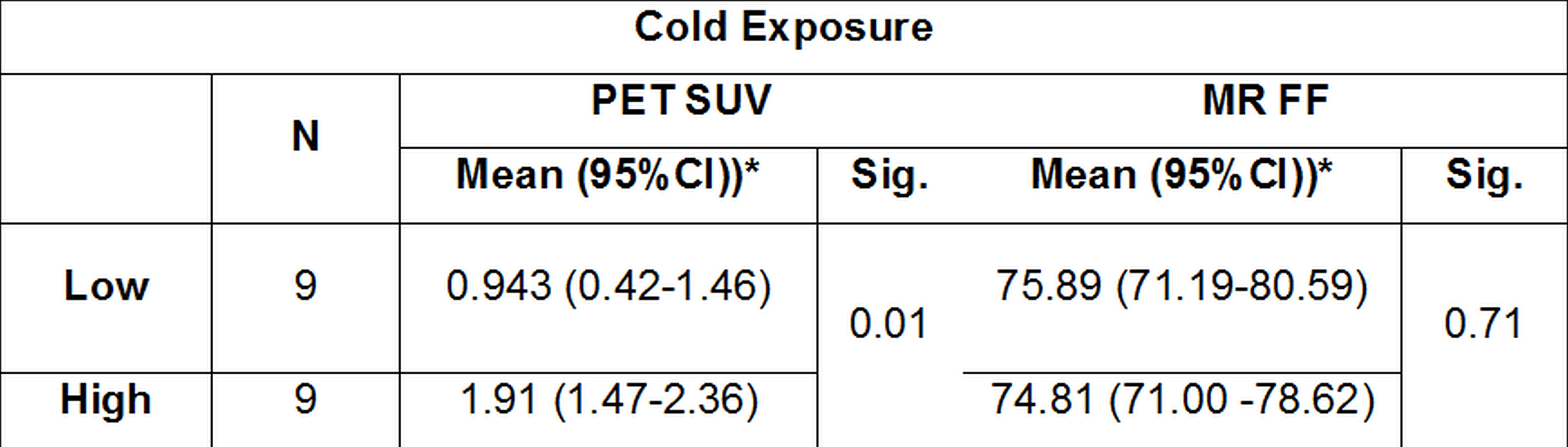
Adaptive thermogenesis after cold exposure was nearly double of that after capsinoids ingestion (207.4 vs 118.7 Cal (p=0.002)). Out of 18 subjects, ten subjects showed the FDG uptake into sBAT during cold exposure. The intervention with oral capsinoids showed minimal or negligible uptake on the supraclavicular depots. Figures 1 and 2 show T2-weighted anatomical, MR, PET and overlaid images of a representative subject with cold exposure and capsinoids intervention respectively. Neither PET nor MRI was capable of distinguishing between high and low responders to capsinoids stimulation, after adjusting for weight and gender (Table 1). The SUV values were significantly higher in higher responders to cold stimulation when compared to low responders, while MRI FF was not significantly different, after adjusting for weight, gender and cold-exposure duration (Table 2). SUV values after cold stimulation were found to be not associated with the duration of cold exposure, while the MRI FF was found to be negatively associated (Figure 3).Discussion and Conclusions
β-adrenergic stimulation in the form of cold exposure and oral capsinoids ingestion has been reported to result in adaptive thermogenesis[1]. The primary aim of this study was to compare PET and MRI for distinguishing high and low responders to the above two stimulants. We found that adaptive thermogenesis after capsinoids ingestion was too low to be detected by either modalities, while PET was successful in identifying high responders to cold stimulation. Unlike PET, MRI-FF seems to be strongly influenced by the duration of cold exposure. Longer durations of cold exposures may be required if MRI is to be used as an alternative to PET for identifying high responders to cold stimulation.Acknowledgements
This research is supported by the Singapore Ministry of Health’s National Medical Research Council under its Clinician Scientist Award Scheme (NMRC/CSA-INV0003/2015).References
- McCallister A, et al. A pilot study on the correlation between fat fraction values and glucose uptake values in supraclavicular fat by simultaneous PET/MRI. Magn Reson Med 2017;78:1922–1932.
- Bhanu Prakash KN, et al. Segmentation and characterization of interscapular brown adipose tissue in rats by multi-parametric magnetic resonance imaging. MAGMA 2016;29:277–86.
- Cinti S. Between brown and white: novel aspects of adipocyte differentiation. Ann Med 2011; 43(2), 104-115.
- Yoneshiro T, et al. Nonpungent capsaicin analogs (capsinoids) increase energy expenditure through the activation of brown adipose tissue in humans. Am J Clin Nutr 2012;95:845–50.
- Hu H, et al. Comparison of brown and white adipose tissues in infants and children with chemical‐shift‐encoded water‐fat MRI. J Magn Reson Imaging 2013;38:885–96.
- Hu HH, et al. Characterization of human brown adipose tissue by chemical-shift water-fat MRI. AJR Am J of Roentgenol 2013;200:177.
- Ren J, et al. Composition of adipose tissue and marrow fat in humans by 1H NMR at 7 Tesla. J Lipid Res 2008;49:2055-2062.
- Schneider CA, et al. NIH Image to ImageJ: 25 years of image analysis. Nat Methods 2012; 9(7):671–675.
- Paul A, et al. User-guided 3D active contour segmentation of anatomical structures: Significantly improved efficiency and reliability. Neuroimage 2006; 1;31(3):1116-28.
- Goh HJ, et al. Gross and relative energy cost of domestic household activities in Asian men. Eur J Clin Nutr 2016; 70(12):1414-1419.
Figures