2499
Magnetic Resonance Imaging and Spectroscopic Investigation of interscapular BAT and Skeletal Muscle IMCL in High Intensity Exercise Trained Rats1Laboratory of Molecular Imaging, Singapore Bioimaging Consortium, Agency for Science, Technology and Research (A*STAR), Singapore, Singapore, 2Mitochondrial Physiology and Metabolism Lab, Department of Physiology, National University of Singapore, Singapore, Singapore
Synopsis
There is a large interest in developing non-pharmacological approaches such as exercise and nutritional compounds for activating BAT to improve metabolic health. In this study, we have investigated the effect of high intensity exercise on interscapular BAT and Intramyocellular lipids (IMCL) from skeletal muscle of rats. Exercise-induced adrenergic receptor stimulation improves quality of iBAT by remodeling of WAT into beige fat and improved mitochondrial fatty acid oxidation. Skeletal muscle IMCL also reduced with exercise along with increased PGC-1α expression due to energy expenditure.
Purpose
To investigate the quality of i-BAT and skeletal muscle IMCL with high intensity exercise intervention.Introduction
Brown adipose tissue (BAT) is metabolically active and plays an important role in maintaining the body energy homeostasis through the adaptive thermogenesis. Recent studies have shown that exercise can stimulate BAT activity[1]. There is a large interest in developing non-pharmacological approaches such as exercise and nutritional compounds for activating BAT to improve metabolic health[2, 3]. In this study, we have investigated the effect of high intensity exercise on interscapular BAT and Intramyocellular lipids (IMCL) from skeletal muscle of rats.Methods
All protocols were in compliance and approved by institutional animal care and use committee. Male Wistar rats (n=12) were fed with normal chow diet (CD) and randomized into exercise (n=6) and non-exercise (n=6) groups. Imaging experiments were performed before and after exercise intervention. Animal treadmill (Columbus-1055SRM-E54 Exer-3/6-Dual) was utilized for exercise interventions and rats were habituated to exercise activity for one week, before subjecting them to high-intensity exercise at the rate of 24 m/min for 45 min/day for 4 weeks. Fat fraction (FF) from iBAT and skeletal muscle IMCL (tibialis anterior) were measured before and after 4 weeks of exercise intervention. MR imaging experiments were performed using a 7T Bruker Clinscan MRI/MRS scanner using a 72 mm volume resonator for RF transmit in combination with 20 mm receive coil. Dixon imaging was performed with FOV 55 mm × 55 mm, matrix size 256 × 256, in-plane resolution 0.214 μm x 0.214 μm, slice thickness 1 mm, TR 8 ms, averages 1, flip angle 8°, echo bandwidths of 1090 and 1500 Hz/pixel, with out-of-phase (1.0 ms) and in-phase (2.5 ms) echo times. Localized PRESS measurements were performed in tibialis anterior muscle with a voxel size of 27 mm3, TR 4 s, TE 13 ms, 2048 complex points, Averages 128 and spectral width of 3500 Hz. Spectra were analyzed using LC Model[4. After terminal experiments, total RNA was isolated from the iBAT and muscle tissues using RNeasy Lipid Tissue Mini Kit (Qiagen 74804) and cDNA conversion using a revertAid H minus first strand cDNA synthesis kit (Thermo Scientific k1632) with oligo d(T) 18 primer according to the manufacturer’s instructions. Real-time qPCR, cDNA samples were analyzed in duplicate using the SYBR Green PCR Master Mix reagent kit (Applied Biosystems 4367659) on a StepOnePlus Real-Time PCR System (Applied Biosystems). Relative mRNA levels were calculated and normalized to 36B4 Forward (TTCCCACTGGCTGAAAAGGT) reverse (GCCGCAGCCGCAAATGC) and GAPDH Forward (TGAACGGGAAGC TCACTG) Reverse (GCTTCACCACCTTCTTGATG) used as an endogenous control gene. The primer sequences used for the UCP1 (ATCTTCTCAGCCGGCGTTTC), CPT-1 acyl group of a long-chain fatty acyl-CoA from coenzyme A to l-carnitine forward (CCGAGCTCAGTGAGGACCTA) reverse (ATCTGTTTGAGGGCTTCGT and CTTGGATCTGAAGGCGGACTTT, PGC-1α forward (TGC CAT TGT TAA GAC CGA G) reverse (GGT CAT TTG GTG ACT CTG G).Results and Discussion
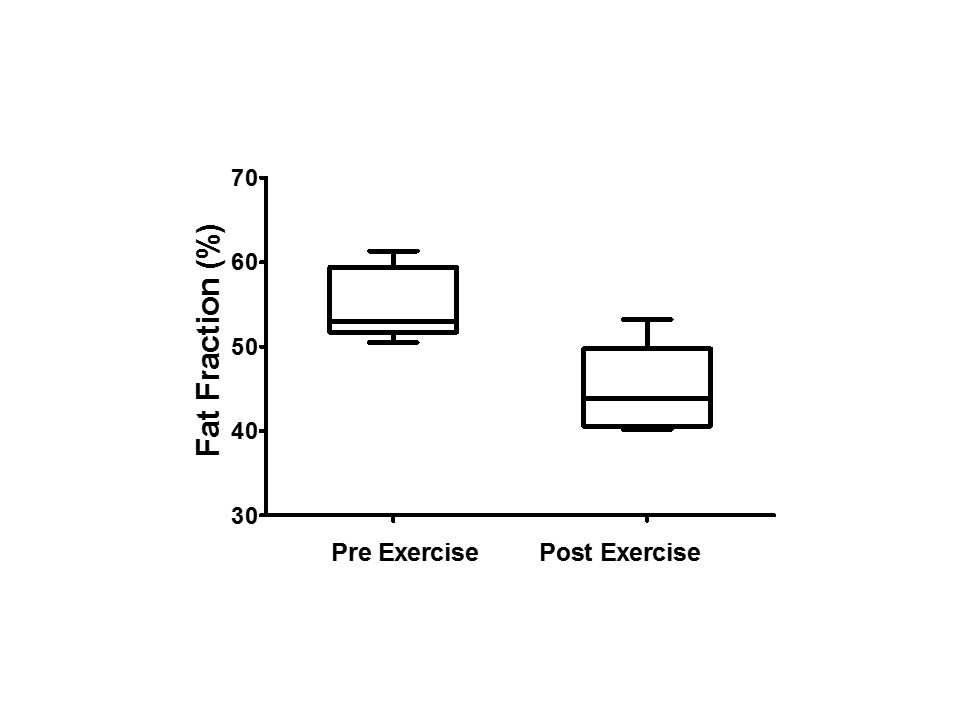
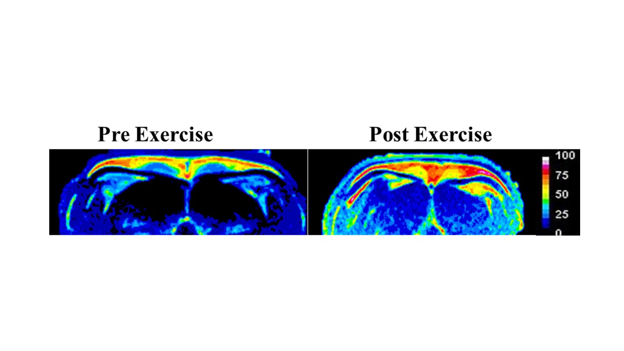
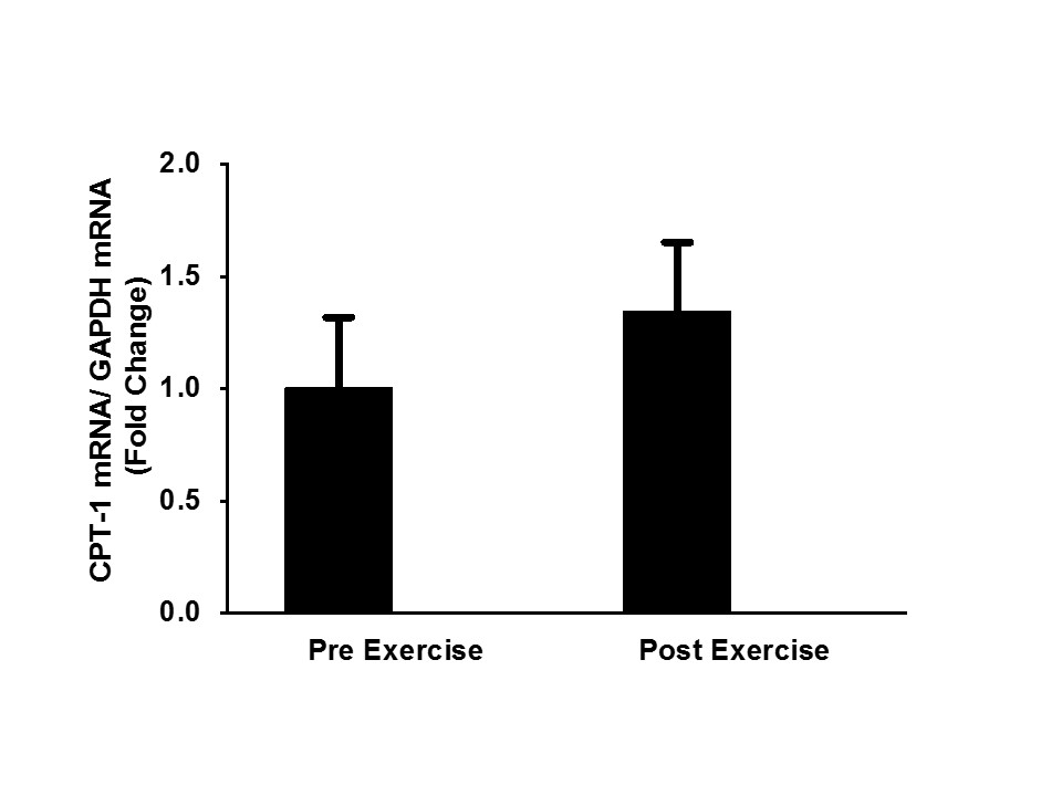
Exercise stimulates sympathetic nervous system and activates BAT to increase thermogenic capacity[1].The fat fraction reduced from 55.03 ± 1.7 % to 44.09 ± 2.1 % after exercise intervention (P<0.05) suggesting remodeling of white adipose tissue into beige fat. The reduction in fat fraction is associated with increased energy expenditure by exercise (Figure 1 and 2). Figure 3 shows the increased expression of carnitine palmitoyltransferase-1 in exercise trained group suggesting improved mitochondrial long chain fatty acid oxidation[5]. Figure 4 shows the IMCL levels of both exercise and sedentary groups. The exercise group shows reduction in IMCL due to increased energy expenditure. Figure 5 shows two fold increase in the PGC-1α expression in exercise group suggesting improved mitochondrial biogenesis due to exercise training[6].Conclusions
Exercise-induced adrenergic receptor stimulation improves quality of iBAT by remodeling of WAT into beige fat and improved mitochondrial fatty acid oxidation. Skeletal muscle IMCL also reduced with exercise along with increased PGC-1α expression due to energy expenditure. Our results show that exercise can improve the metabolic health by increasing the quality of brown fat and also reduction of IMCL in skeletal muscle.Acknowledgements
No acknowledgement found.References
1. R. De Matteis et al., Nutrition, Metabolism & Cardiovascular Diseases 2013: 23: 582-590.
2. Wallberg-Henriksson H et al., Nat Rev Endocrinol 2015;11(4):198-200.
3. Leslie P. Kozak et al., Cell Metabolism:2010:11:263-267.
4. Provencher et al., NMR Biomed 2001: 14: 260.
5. María Calderon-Dominguez et al., Adipocyte 2016:5:2: 98-118.
6. Chounghun
Kang et al., Ann. N.Y. Acad. Sci 2012:1271:110-117.
Figures