2491
Intravoxel incoherent motion-diffusion weighted imaging (IVIM-DWI) parameters distinguish kidney allografts with delayed graft functionEyesha Hashim1, Darren Yuen2,3, General Leung1,4, and Anish Kirpalani1,4
1Medical Imaging, St. Michael's Hospital, Toronto, ON, Canada, 2Nephrology, St. Michael's Hospital, Toronto, ON, Canada, 3Keenan Research Centre for Biomedical Science, St. Michael's Hospital, Toronto, ON, Canada, 4Li Ka Shing Knowledge Institute, St. Michael's Hospital, Toronto, ON, Canada
Synopsis
Delayed graft function (DGF) complicates 21-36% of all deceased donor kidney transplants, and leads to early inpatient post-transplant dialysis, higher risk of graft failure and death. In this abstract, we show that IVIM-derived flow (f)-fraction, is significantly different in kidney allografts exhibiting DGF compared to those that do not develop DGF. Furthermore, f fraction shows a significant negative correlation with time to recovery and a positive trend with renal function at 3 months post-transplant as measured with eGFR.
Introduction
While kidney transplantation has revolutionized the care of patients with end-stage kidney disease, the use of kidneys from older donors and donors after cardiac arrest has dramatically increased the frequency of dysfunction of transplanted kidneys immediately after implantation. This “delayed graft function” (DGF), defined as the need for dialysis within the first week post-transplantation1, is estimated to complicate 21-36% of all deceased donor kidney transplants 2, and its incidence continues to rise. Importantly, DGF is not just a short term problem, but is also associated with a higher risk of later graft failure 3. Moreover, even if graft function persists, DGF is also associated with a higher rate of death 4. Currently, no clinical tools exist that accurately predict the time to, and degree of, kidney function recovery following DGF. DGF thus leads to longer hospitalization stays, patient anxiety, and more complex discharge planning and outpatient care. In this abstract, we investigated: (1) whether intravoxel incoherent motion (IVIM)-derived flow (f)-fraction is different in kidney allografts exhibiting DGF compared to those that do not develop DGF, and (2) whether f fraction correlates with time to recovery as measured by the time to come off of dialysis post-transplantation and with resumption of normal kidney function at 3 months post-transplant as measured with estimated glomerular filtration rate (eGFR)Methods
This prospective study was approved by our institutional review board. Twenty (20) kidney transplant inpatients, 10 with DGF and 10 with initial graft function, at our hospital consented to be part of this study. The clinical data included a record of dialysis, blood tests during hospital stay and at 3-months post-transplant. IVIM-DWI was performed within 10 days post-transplant, on a 3.0 T MRI (Siemens Skyra) scanner with the following parameters: single shot EPI readout, TR / TE = 3500 / 68 ms, 5 x 5 mm thick slices for each of seven b values: 0, 50, 100, 300, 600, 800 and 1000 s/mm2, NEX=2 for images with b >100 s/mm2, acquisition matrix=126x106 reconstructed to 256x256. The images were post-processed using a bi-exponential IVIM equation5 in Matlab6 to calculate f-fraction on a pixel-by-pixel bases. The regions of interest (ROIs) were drawn to represent cortex, medulla and the entire renal parenchyma on 3-5 slices. The average of all pixels in the ROIs was calculated to obtain cortical, medullar and parenchymal values for f-fraction. The person processing the images was blind to the DGF status and clinical data of patients. eGFR was calculated from serum creatinine values using the Modification of Diet in Renal Disease formula7 3-months post-transplant, a time when most transplant kidneys will have reached their maximum recovered function. Time to recovery was defined as the number of days post-transplant until the last dialysis session was needed. Student’s t-test was used to compare the parameters between the two groups. Spearman’s correlation analysis was performed to assess if f-fraction correlated with time to recovery and resumption of normal kidney function at 3 months post-transplant.Results
We found significantly lower f-fraction in the DGF group compared to patients that do not develop DGF for f-fraction measured in both the cortex and medulla (Figure 1). We also found a significant negative correlation between f-fraction and time to recovery (Figure 2) in the cortex, the medulla and entire renal parenchyma with Spearman’s correlation coefficients of -0.69 (p=0.02), -0.69 (p=0.02) and -0.74 (p=0.01) respectively. Finally, we found a positive trend between f-fraction and eGFR at 3 months post-Transplant in the cortex, the medulla and entire renal parenchyma (Figure 3).Discussion and Conclusion
While other reports, using multiple imaging modalities including DWI, have demonstrated lower perfusion in transplant kidneys with DGF8,9, our results are the first to also show that MRI-derived f-fraction correlates with time to recovery post -DGF. In this small pilot study, we also demonstrate a positive trend between f-fraction and kidney function (as measured by eGFR) at 3 months post-transplant. Our findings suggest that IVIM-DWI could be used to predict time to recovery and thus aid in planning of care post-transplant.Acknowledgements
The authors wish to thank the research MRI technicians at our hospital, Cindy Hamid and Anthony Sheen, for their help in performing the imaging.References
1. Perico, N., Cattaneo, D., Sayegh, M.H. & Remuzzi, G. Delayed graft function in kidney transplantation. Lancet 364, 1814-1827 (2004). 2. Bronzatto, E.J., da Silva Quadros, K.R., Santos, R.L., Alves-Filho, G. & Mazzali, M. Delayed graft function in renal transplant recipients: risk factors and impact on 1-year graft function: a single center analysis. Transplant Proc 41, 849-851 (2009). 3. Giral-Classe, M., et al. Delayed graft function of more than six days strongly decreases long-term survival of transplanted kidneys. Kidney Int 54, 972-978 (1998). 4. Tapiawala, S.N., et al. Delayed graft function and the risk for death with a functioning graft. Journal of the American Society of Nephrology : JASN 21, 153-161 (2010). 5. Le Bihan, D., et al. Separation of diffusion and perfusion in intravoxel incoherent motion MR imaging. Radiology 168, 497-505 (1988). 6. The MathWorks Inc. (2014): MATLAB. (Natick, Massachusetts.). 7. Levey, A.S., et al. A more accurate method to estimate glomerular filtration rate from serum creatinine: a new prediction equation. Modification of Diet in Renal Disease Study Group. Ann Intern Med 130, 461-470 (1999). 8. Ren, T., et al. Evaluation of renal allografts function early after transplantation using intravoxel incoherent motion and arterial spin labeling MRI. Magn Reson Imaging 34, 908-914 (2016). 9. Hueper, K., et al. Diffusion-Weighted imaging and diffusion tensor imaging detect delayed graft function and correlate with allograft fibrosis in patients early after kidney transplantation. J Magn Reson Imaging 44, 112-121 (2016).Figures
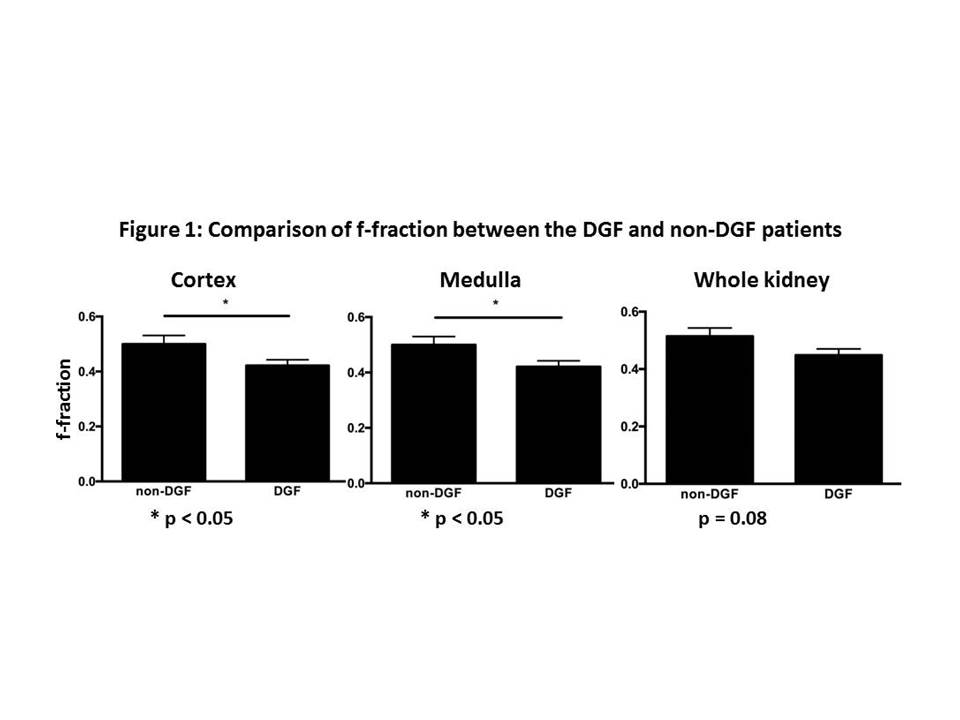
Figure 1: Comparison of f-fraction between DGF and non-DGF groups in the cortex, medulla and entire renal parenchyma (whole kidney).* represents the Student's t-test p < 0.05.
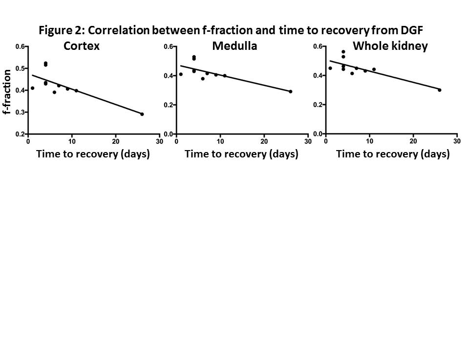
Figure 2: Correlation
between f-fraction and time to recovery in the DGF group in the cortex,
medulla and entire renal parenchyma (whole kidney).
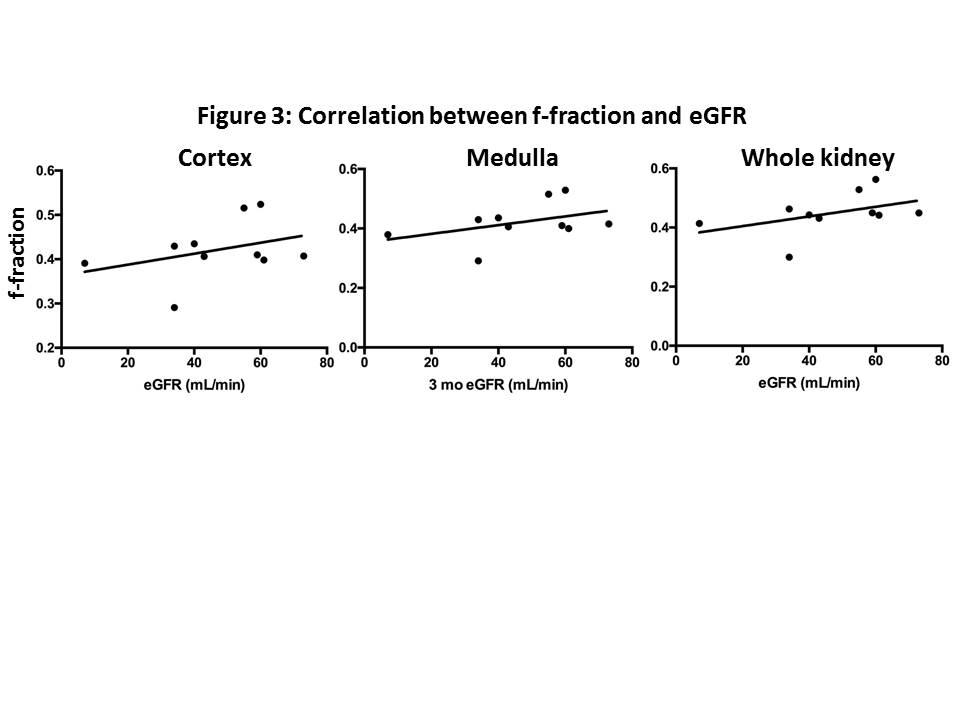
Figure 3: Correlation between f-fraction and eGFR ( a measure of normal renal function) at 3 months post-transplant in the DGF group.