2489
Case report: Three-dimensional visualization of the normal human perirectal muscle with diffusion tensor imaging (DTI)1Department of Diagnostic Imaging and Nuclear Medicine, Kyoto University Graduate School of Medicine, Kyoto, Japan, 2Human Brain Research Center, Kyoto University Graduate School of Medicine, Kyoto, Japan, 3Division of Gastrointestinal Surgery, Department of Surgery, Kyoto University Graduate School of Medicine, Kyoto, Japan, 4Siemens Healthcare K. K., Tokyo, Japan
Synopsis
Diffusion tensor imaging (DTI) can provide the directionality of water diffusion in tissues, informing on its underlying microstructures and microdynamics. There has been no previous report on the visualization of anterior portion of the longitudinal anal muscle (aLAM). In this case study, we present the 3D visualization of the aLAM in normal male subjects with DTI. By adjusted parameters for DTI sequence, we could successfully visualize thin smooth muscle layer of the rectum. This technique could be useful when planning operation for rectal and anal diseases.
Introduction
Transanal operation for lower rectal cancer or other rectal diseases has been developed recently [1]. In this procedure, we should understand the architecture of the muscles or layers around the rectum and anal canal. The topography of longitudinal anal muscle (LAM) has been reported in recent anatomical study [2]. We revealed the difference of the topography between superior and inferior part of anterior portion of the longitudinal anal muscle (aLAM) from our anatomical study of perirectal space on cadaveric specimens [3]. Diffusion tensor imaging (DTI) can non-invasively provide the directionality of water diffusion in tissues in vivo, informing on its underlying microstructures and microdynamics [4,5,6]. The visualization of pelvic floor or anal canal in normal human has been reported [7]. However, detailed depiction of aLAM has not been reported. We performed MR-DTI with adjusting several parameters in DTI sequence and could depict aLAM more precisely. The purpose of this study was to evaluate feasibility of DTI technique for the visualization and depiction of aLAM in normal human.Methods
MRI protocol
DWI was performed to three healthy male volunteers on a 3T clinical MRI scanner (MAGNETOM Skyra; Siemens Medical Solutions, Erlangen, Germany) using a six-channel body matrix coil. After obtaining three-plane localizing images, DWI was acquired in the axial plane, encompassing the length of the anal canal and prostate. The parameters were as follows: (DWI) TR/TE, 3300/43 ms; matrix size, 134×134 (268×268 with interpolation); FOV, 200×200 mm; acquisition bandwidth, 1866 Hz/pixel; b-values, 0 and 400 s/mm2; diffusion direction, 64 orientations plus one null; number of averages, 2; number of slices, 24; parallel imaging factor, 2; phase partial Fourier, 6/8; slice inter-leave, on. Nominal voxel size was 0.75×0.75×5 mm with no inter-slice gap. For fat suppression, the CHESS pulse and gradient reversal technique [8] was used. The acquisition time for DWI was 10 minutes 47 seconds.
Data processing and visualization
DTI datasets were processed using Diffusion Tool Kit (http://www.trackvis.org/dtk), and pelvic streamline tracking procedure was carried out using TrackVis (http://www.trackvis.org) [9,10].
First, an apparent diffusion coefficient (ADC) map and fractional anisotropy (FA) map were calculated from DWI datasets using Diffusion Tool Kit (http://www.trackvis.org/dtk). ADC was calculated on a voxel-by-voxel basis as follows:
ADC=1/b × -ln(S/S0),
where S0 and S are the signal intensities of each voxel obtained with b-values of 0 and 400 s/mm2, respectively. FA was calculated using the following formula:
FA=√½[√{(λ1-λ2)2+(λ1-λ3)2+(λ2-λ3)2} / √(λ12+λ22+λ32)],
where λ1, λ2, and λ3 correspond to three eigenvalues of the diffusion tensor.
Second, fiber tracking was performed by using a FACT algorithm with the angle threshold of 35° using the same software (Diffusion Toolkit) [11].
For the next step, a three-dimensional volume of interest (VOI) was carefully drawn by one of the authors on the DWI data with a b-value of 0 s/mm2 by using TrackVis. VOIs for the aLAM, epithelial/subepithelial layer of the rectum and anal canal, internal anal sphincter, external anal sphincter, puborectalis muscle, superficial transverse perineal muscle, and gluteus maximus muscle were drawn separately. Tracked data of the fibers were visualized using TrackVis.
Results
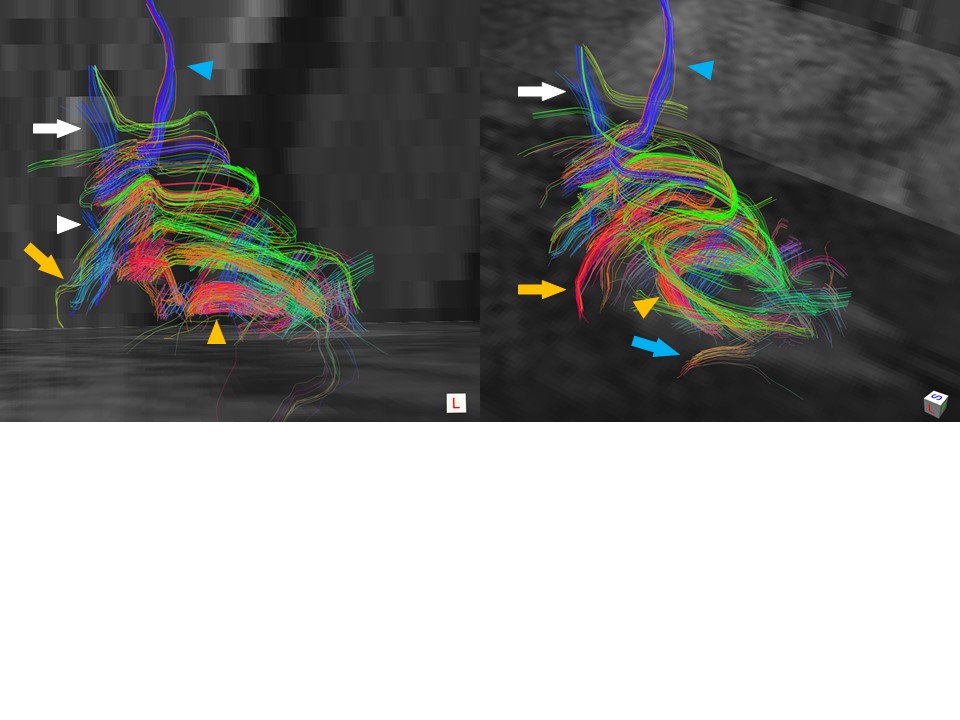
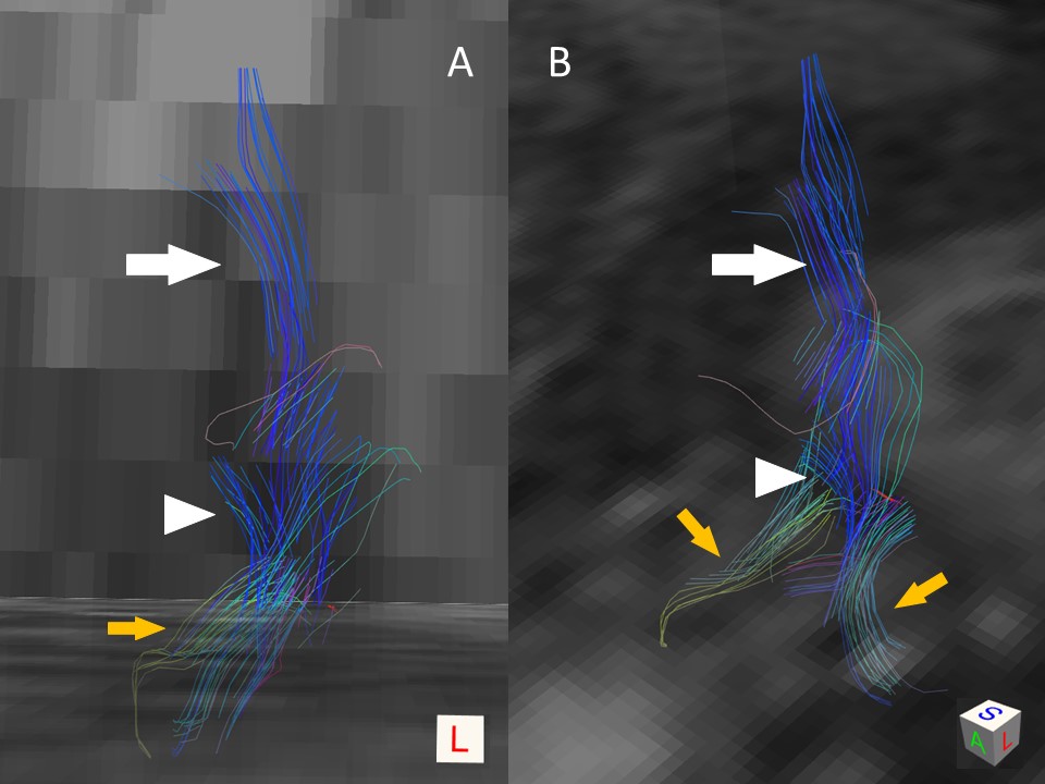
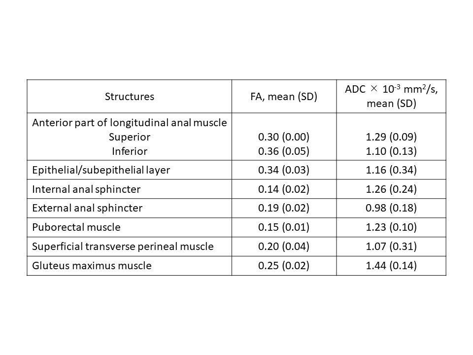
aLAM, epithelial/subepithelial layer of the rectum and anal canal, internal anal sphincter, external anal sphincter, puborectalis muscle, and superficial transverse perineal muscle were visualized in 3D (Fig.1). The stream of both superior and inferior part of aLAM fiber ended at the level of bulbocavernosus muscle (Fig.2). Superior aLAM was depicted in all three cases, while inferior aLAM was depicted in two cases. FA and ADC differed across each structure (Table.1).Conclusion
In this study, DTI of aLAM was performed, and the aLAM was visualized and depicted.
Precise visualization and segmentation of perirectal muscle using DTI can provide better understanding of the anatomy around rectum and anal canal, which is important for anal and rectal operation.
Acknowledgements
The authors have no potential conflict of interest related to this presentation.References
1. Tuech JJ, Bridoux V, Kianifard B, Schwarz L, Tsiliividis B, Huet E, Michot F. Natural orifice total mesorectal excision using transanal port laparoscopic assistance. Eur J Surg Oncol 2011; 37(4): 334-335.
2. Macchi V, Porzionato A, Stecco C, Vigato E, Parenti A, De Caro R. Histo-topographic study of the longitudinal anal muscle. Clin Anat 2008; 21(5): 447-452.
3. Okada T, et al. The 30th annual meeting of the Kinki Society for Endoscopic Surgery. Sept, 2017.
4. Basser PF, Pierpaoli C. Microstructural and physiological features of tissues elucidated by quantitative-diffusion-tensor MRI. J Magn Reson B 1996; 111(3): 209-219.
5. Le Bihan D, Breton E, Lallemand D, Aubin ML, Vingnaud J, Laval-Jeantet M. Separation of diffusion and perfusion in intravoxel incoherent motion MR imaging. Radiology 1988; 168(2): 497-505.
6. Fujimoto K, Kido A, Okada T, Uchikoshi M, Togashi K. Diffusion tensor imaging (DTI) of the normal human uterus in vivo at 3 tesla: Comparison of DTI parameters in the different uterine layers. J Magn Reson Image 2013; 38(6): 1494-1500.
7. Goh V, Tam E, Taylor NJ, Stirling JJ, Simcock IC, Jones RG, Padhani AR. Diffusion tensor imaging of the anal canal at 3 tesla: Feasibility and reproducibility of anisotropy measures. J Magn Reson Image 2012; 35(4): 820-826.
8. Park HW, Kim DJ, Cho ZH. Gradient reversal echnique and its applications to chemical-shift-related NMR imaging. Magn Reson Med 1987; 4(6): 526-536.
9. Pieper S, Lorensen B, Schroeder W, Kikinis R. The NA-MIC Kit: ITK, VTK, pipelines, grids and 3D slicer as an open platform for the medical image computing community. I S Biomed Imaging 2006; 698-701.
10. Wang R, Benner T, Sorensen AG, Wedeen VJ. Diffusion toolkit: a software package for diffusion imaging data processing and tractography. Proc Intl Soc Mag Reson Med 2007; 15: 3720.
11. Mori S, Crain BJ, Chacko VP, van Zijl PC. Three-dimensional tracking of axonal projections in the brain by magnetic resonance imaging. Ann Neurol 1999; 45(2): 265-269.
Figures