2482
Semi-automatic method for generating multiplanar reformatting views of MR post-contrast T1-weighted images for visualizing and assessing pediatric Crohn’s disease1Radiology, Boston Childrens Hospital and Harvard Medical School, Boston, MA, United States
Synopsis
In this proposed study, we aim to develop a semi-automated method for generating multiplanar reformatting images (MPR) of pediatric Crohn’s disease (pCD) segments from T1-weighted post-contrast MR image data. We demonstrate that this method can efficiently visualize and assess this disease. Importantly, the centerline length can be used as a reliable measure of the extent of disease. Moreover, the MPR image can be used as a platform for intestinal wall segmentation and for more accurate depiction of luminal narrowing. We also expect such MPR views to be used as a unified parametric platform for evaluating disease progression in follow-up scans.
INTRODUCTION
To develop the most optimal plan-of-care for patients with pCD, disease progression and severity must be assessed early on in the course of this illness. Moreover, a timely and accurate diagnosis has important prognostic implications. The use of magnetic resonance imaging (MRI) for assessing pCD has increased dramatically, especially as we strive to spare children the potentially harmful effects of radiation when imaged with CT. Wall thickness, wall enhancement, length of the inflamed segment, and luminal narrowing are important biomarkers of disease activity1. However, due to the curved structure of the bowel, these features are often difficult to measure in the coronal plane acquisition. In addition, the lack of spatial alignment in follow-up scans makes it challenging to compare similar regions.METHOD
Our method entails centerline extraction as well as generation of MPR images. In advance of this analysis, however, we resampled the images to an isotropic resolution. Throughout the processing, we used a dedicated, interactive MATLAB editing tool to easily adds/correct the contours in the coronal images and fix the locations of the centerline points.
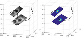
Centerline extraction: For centerline extraction, we perform the following stages: First, we segment the bowel's lumen in the coronal images by manually positioning a few seed points in its internal region and then detecting the connected lumen region. Next, we extract the skeleton of the implicit surface enclosed by the lumen contours using a method similar to 2. We start with non-ordered set of the lumen’s central points from the coronal image slices. We then search for the center line's start and end points by locating the two points with the maximal geodesic distance between them. Next, we generate an implicit surface from the lumen contours and calculate the shortest distance map from the lumen's boundary into its internal region. Last, we use the boundary distance map as a speed image for finding the shortest path between the centerline's start and end points (Figure4). We back up the method with an editing tool of three synchronized screens of coronal, sagittal and axial views crossing an interactively selected point of interest to add centerline points.
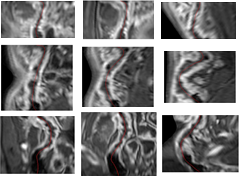
Generation of Multiplanar Reformatting Views: For curved MPR views, we find a plane that transverses through the centerline's start and end points and a third point on the centerline, selected interactively by the operator using the mouse scroller. The curve is then projected in the direction normal to the plane. After performing arc-length parameterization3 to the projected centerline, each row in the curved MPR image is obtained by interpolating the values vertical to the projected curve points. For straight MPR views, we use the Frenet–Serret formulas4 to find the local axis system along the centerline. We use the Savitsky-Golay filtering5 to smooth the curve and the vector’s components along the centerline. We then interpolate the image values in the normal planes to generate cross-sectional views.
RESULTS
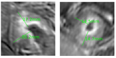
We have generated MPR views of CD segments in 12 pediatric patients. The T1-weighted images were scanned in coronal planes with average resolution of (0.7x0.7x2) mm after contrast injection. The CD segment length range was [4.3, 31.5] cm. In preliminary measurements, we obtained an under estimation of 15%, on average, when using a series of points to estimate the abnormal segment length in the coronal planes compared to the centerline's length. An over estimation of 20-40% in stricture wall thickness was obtained when measuring it in the coronal plane acquisition (Figure 3).
DISCUSSION
Although MPR views are routinely used for analyzing the luminal narrowing of arteries and the surface of the large bowel, to the best of our knowledge, there are no easily mastered tools for analyzing MR images of the small bowel. In this proposed study, we will show that the centerline of CD segments as well as the generation of MPR views can be used to assess and quantitatively evaluate features of pCD. The length of the centerline can also be used to measure the bowel's segment size in a more reliable fashion. The thickness of the wall is more accurate when it is measured in the normal planes (Figure3). Finally, the lumen is better demonstrated in the curved MPR views (Figure1). When starting the centerline in the same anatomical position in two follow-up scans (e.g., the ileocecal valve), the change in disease markers indicating response-to-therapy is likely to be analyzed more effectively using MPR views.CONCLUSION
MPR views can be used as an effective platform for quantitatively assessing bowel segments in pediatric Crohn’s disease.Acknowledgements
No acknowledgement found.References
1. Rimola J, Rodríguez S, García-Bosch O, Ordás I, Ayala E, Aceituno M, Pellisé M, Ayuso C, Ricart E, Donoso L, Panés J. Magnetic resonance for assessment of disease activity and severity in ileocolonic Crohn’s disease. Gut. 2009 Aug 1;58(8):1113-20.
2. Van Uitert R, Bitter I. Subvoxel precise skeletons of volumetric data based on fast marching methods. Medical physics. 2007 Feb 1;34(2):627-38.
3. Willmore TJ. An introduction to differential geometry. Courier Corporation; 2013 May 13.
4. Weatherburn CE. Differential geometry of three dimensions. Cambridge University Press; 2016 Apr 15.
5. Orfanidis SJ. Introduction to signal processing. Prentice-Hall, Inc.; 1995 Nov 1.
Figures