2466
UTE-SENCEFUL: high resolution 3D ventilation weighted maps1Department of Diagnostic and Interventional Radiology, University Hospital Wurzburg, Wurzburg, Germany
Synopsis
In this work we present a method to assess lung ventilation in 3D by combining Self-gated Non-Contrast-enhanced Functional Lung MRI (SENCEFUL) with an ultra-short echo time (UTE) acquisition and a 3D image registration technique. Ventilation weighted maps were generated and the quantitative ventilation value for a healthy volunteer was assessed. Lung ventilation and image quality were compared between the new UTE-SENCEFUL and the standard 2D-SENCEFUL methods. UTE-SENCEFUL was able to present a 3D reconstruction of the breathing cycle, 3D ventilation weighted maps with high resolution and quantitative ventilation values in agreement with the literature.
Introduction
Self-gated Non-Contrast-enhanced Functional Lung MRI (SENCEFUL) is a technique able to assess ventilation and perfusion in free-breathing 1,2. Its standard implementation is based on a custom 2D-FLASH sequence, where the direct current (DC) signal is sampled after each readout1. However, 2D-FLASH suffers from the T2* effect in the lung parenchyma and it does not offer real 3D coverage. One solution to assess ventilation in the whole lung is to acquire consecutive parallel 2D slices across the lung; but long measurement times (between 30 and 45 minutes) and poor resolution in the slice direction are limiting factors for application in clinical routine. To better account for the T2* effect, a 3D-UTE sequence with a koosh ball trajectory has been previously implemented in SENCEFUL MRI 3. High resolution ventilation maps were then generated using a quasi-random sampling scheme combined with the 3D-UTE acquisition, which resulted in a better filling of the k-space and shorter measurement times4. Despite the improvement in image quality (especially of the morphological images), blurring and ventilation artifacts were still common due to the use of a 2D image registration technique, which was not able to account for in-plane motion and ultimately prevented the generation of ventilation maps in 3D. Furthermore, image quality was corrupted by gradient delays and distortions, which were ignored in our reconstruction so far. Thus, to acquire ventilation weighted maps in 3D and improve overall image quality, this work presents a 3D image registration technique and the correction of gradient deviations in the UTE-SENCEFUL framework.Methods
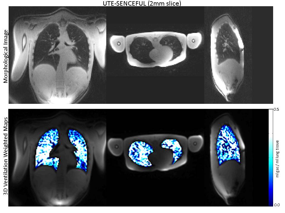
A 3D-UTE sequence with a nonselective RF pulse and koosh ball quasi-random sampling order was developed for a 3 T MR scanner (MAGNETOM Prisma, Siemens Healthcare, Erlangen) equipped with a 32-channel coil-array. Measurements were performed in a healthy volunteer in tidal breathing and supine position. The following parameters were adjusted: TE = 0.03 ms; TR = 1.49ms; flip angle = 2°; FOV = 350x350mm; number of projections = 350000, resolution = 2.7mm x 2.7mm, slice thickness = 2mm. The DC signal from a single coil element, where the breathing motion could be depicted, was chosen for data binning. This signal was filtered and used as navigator for the segmentation of the k-space into eight individual k-spaces, each representing one breathing phase, from expiration to inspiration. Each breathing phase had a sampling density of at least 78% of the Nyquist sampling rate at Kmax. Prior image reconstruction, gradient delays and trajectory errors were correct using the Gradient Impulse Response Function5. Iterative SENSE6 was then applied to determine the fully sampled data. In order to eliminate signal changes caused by motion, all breathing phases were morphed onto a phase of reference using 3D image registration7. Ventilation for the whole the lung was quantified and ventilation weighted maps were generated by comparing the signal changes in inspiration and expiration phases8. For quantification of pulmonary function the lungs were manually segmented. Finally, image quality and quantitative ventilation (QV) values were compared to the standard 2D SENCEFUL technique.Results
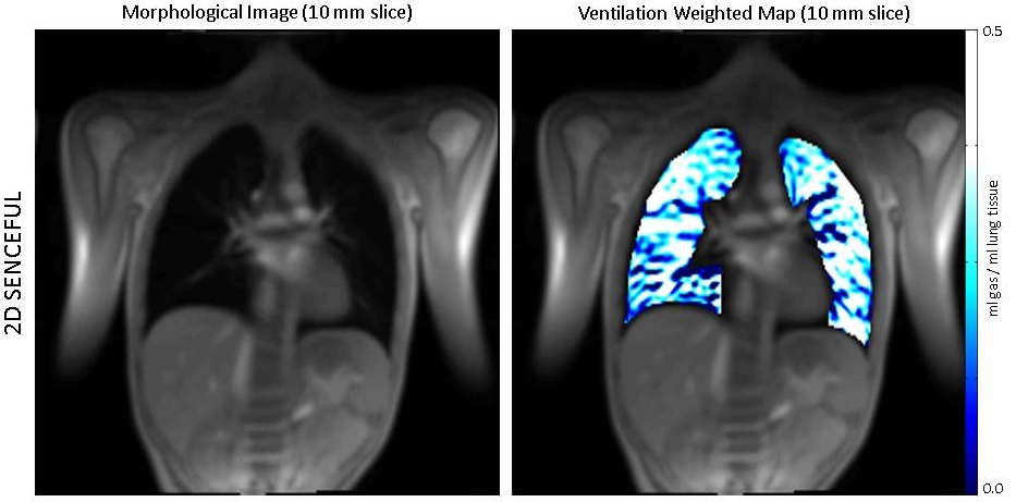
Figure 1 presents the ventilation weighted map generated with the standard 2D SENCEFUL technique and the corresponding morphological image after 2D image registration. In Figure 2 three out of eight breathing phases of the 3D-UTE acquisition without image registration can be observed. In this figure, the different positions of the diaphragm during a breathing cycle can be clearly delineated. The improved SNR allows for the visualization of parenchyma and blood vessels, which were not visible in Figure 1. Figure 3 shows one breathing phase after 3D image registration and the corresponding ventilation weighted maps generated with UTE-SENCEFUL for the coronal, axial and sagittal planes. The average quantitative ventilation values in ml of gas per ml of lung tissue for both UTE-SENCEFUL and 2D SENCEFUL for the coronal plane are respectively: 0.11 ± 0.07 ml/ml and 0.11 ± 0.08 ml/ml.Discussion
The improved implementation of UTE-based SENCEFUL MRI was able to provide high resolution 3D lung images with less artifacts and blurring. The technique allowed for the calculation of 3D ventilation maps, which renders the investigation less prone to partial volume effects and motion artifacts in comparison to the conventional 2D SENCEFUL approach. The average quantitative ventilation value for UTE-SENCEFUL was in agreement with the literature for a healthy volunteer2. Finally, measurement time was reduced from 31.2 to 8.6 minutes for the whole lung. UTE-SENCEFUL thus represents an alternative technique to assess ventilation in the human lung.Acknowledgements
No acknowledgement found.References
1. Fischer A, Weick S, Ritter C. O, et al. SElf-gated Non-Contrast-Enhanced FUnctional Lung imaging (SENCEFUL) using a quasi-random fast low-angle shot (FLASH) sequence and proton MRI. NMR in Biomedicine. 2014.
2. Veldhoen S, Weng A. M., Knapp J, et al. Self-gated Non-Contrast-enhanced Functional Lung MR Imaging for Quantitative Ventilation Assessment in Patients with Cystic Fibrosis. Radiology. 2016.
3. Mendes Pereira L, Wech T, Weng A. M, et al. Self-gated ultra-short echo time lung MRI for quantitative ventilation assessment. Proceedings ISMRM 2017.
4. Mendes Pereira L, Weng A. M, Wech T, et al. UTE-SENCEFUL: high resolution 3D ventilation weighted maps. Proceedings ESMRMB 2017.
5. Stich M, Wech T, Slawig A, et al. Implementation of a Gradient Pre-emphasis Based on the Gradient Impulse Response Function. Proceedings ISMRM 2017.
6. Pruessmann KP, Weiger M, Bornert P, et al. Advances in sensitivity encoding with arbitrary k-space trajectories. Magn Reson Med. 2001.
7. Kroon, D.-J., & Slump, C. H. MRI modalitiy transformation in demon registration. IEEE International Symposium on Biomedical Imaging: From Nano to Macro, 2009.
8. Zapke M, Topf HG, Zenker M, et al. Magnetic resonance lung function - a breakthrough for lung imaging and functional assessment? A phantom study and clinical trial. Respiratory Research. 2006.
Figures