2452
Free-breathing T1-weighted 3D STAR VIBE: versus Thin-Section Computed Tomography for the Assessment of Pulmonary Parenchyma DiseasesZhanli Ren1, Shan Dang2, Yuxin Lei2, Nan Yu2, Yong Yu2, and Taiping He2
1Shaanxi University of Chinese Medicine, Xianyang, China, 2Affiliated Hospital of Shaanxi University of Chinese Medicine, Xianyang, China
Synopsis
Free-breathing T1-weighted 3D star vibe is useful for lung and mediastinum assessment and evaluation of radiological findings for patients with various pulmonary parenchyma diseases.
Purpose
To evaluate the performance of pulmonary MR imaging with free-breathing T1-weighted 3D star vibe in assessment of pulmonary parenchyma diseases,with computed tomography (CT) as the reference standard.Methods
30 consecutive patients (25 males and 5 females: mean age 64 years) with various pulmonary parenchyma lesions were detected on chest thin-section standard-dose CT as well as pulmonary MR imaging with Star Vibe. A 5-point visual scoring system for assessment in lung parenchyma was adopted (1 absent; 2 probably absent; 3 equivocal; 4 probably present; 5 present), including masses(≥2cm), nodules(<2cm), consolidation, patchy shadow, cords focal, mesh shadow, ground glass density, mediastinal lymph nodes, pleural thickening, pleural effusion, emphysema, pulmonary bullae, traction bronchiectasis and so on. Chest CT images of each patient were reviewed by a chest radiologists with 25 years of experience, and the results were used as the reference standard for radiological findings. Then, all images obtained with standard-dose CT and Star VIBE (MRI) were randomized and independently evaluated by two chest radiologists without any information about the results. Receiver operating characteristic analyses were used to compare diagnostic performance.Results
The detection rates of pulmonary masses, consolidation, mediastinal lymph nodes and pleural effusion were 100% with pulmonary MR free-breathing T1-weighted 3D star vibe. The MR imaging can detect pulmonary nodules and pleural thickening with a high rate about 93.7% and 92.9%.The patchy shadow, cords focal, mesh shadow can also be displayed at a rate of 81.8%-88%.The detection rates of ground glass density and traction bronchiectasis was lower than other disease other than emphysema and bullae. The pulmonary MR free-breathing T1-weighted 3D star vibe would miss all emphysema and bullae.Conclusion
Pulmonary MR imaging with free-breathing T1-weighted 3D star vibe is useful for lung and mediastinum assessment and evaluation of radiological findings for patients with various pulmonary parenchyma diseases.Acknowledgements
No acknowledgement found.References
[1] Cieszanowski A, Lisowska A, Dabrowska M, et al. MR Imaging of Pulmonary Nodules: Detection Rate and Accuracy of Size Estimation in Comparison to Computed Tomography[J]. Plos One, 2016, 11(6):e0156272.
[2] Yedururi S, Kang H C, Wei W, et al. Free-breathing radial volumetric interpolated breath-hold examination vs breath-hold cartesian volumetric interpolated breath-hold examination magnetic resonance imaging of the liver at 1.5T[J]. World J Radiol. 2016, 8(7):707-715.
Figures
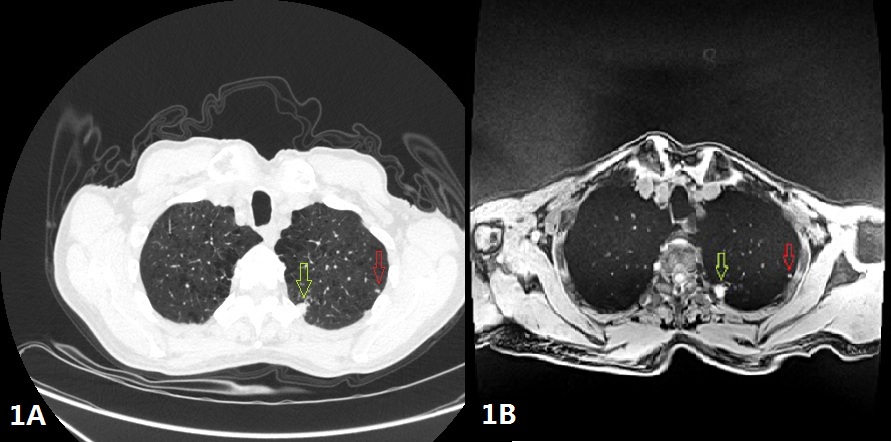
Figure 1A-1B A 59-year–old male with nodules located in the upper lobe of left lung,the size of the nodules were 3mm(Red arrow) and 9mm(,Green arrow),these nodules can be seen on CT(Figure 1A) as well as on Star VIBE(Figure 1B).
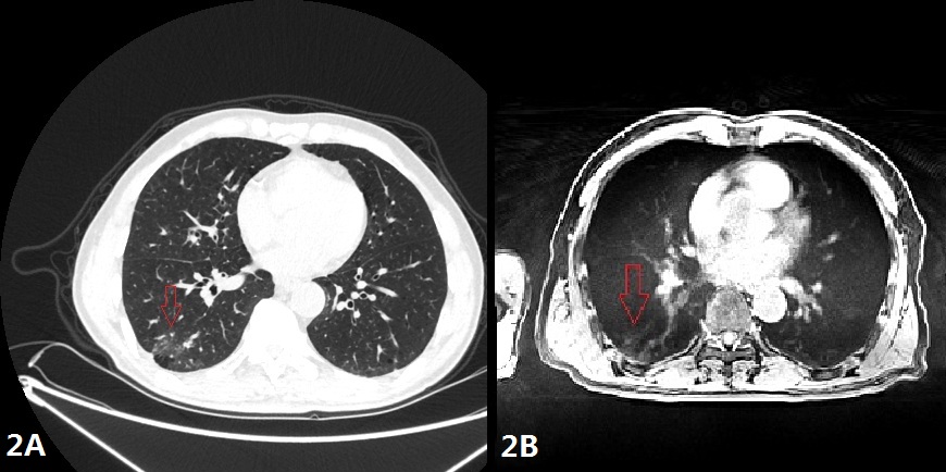
Figure 2A-2B A 51-year–old female with ground glass density(Red arrow),it can be observed on CT(Figure 2A) as well as on Star VIBE(Figure 2B).
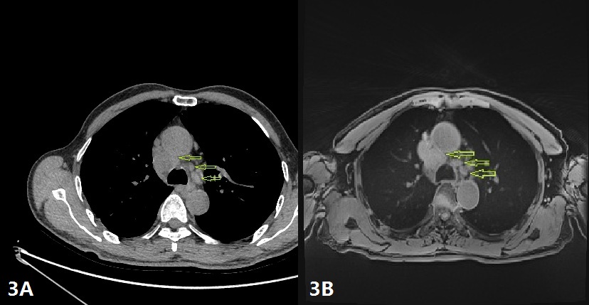
Figure 3A-3B A 28-year–old female with lymph nodes(Green arrow) can be seen in aorta-pulmonary artery window,and all the lymph nodes can be showed clearly on CT (Figure 3A) as well as Star VIBE(Figure 3B)
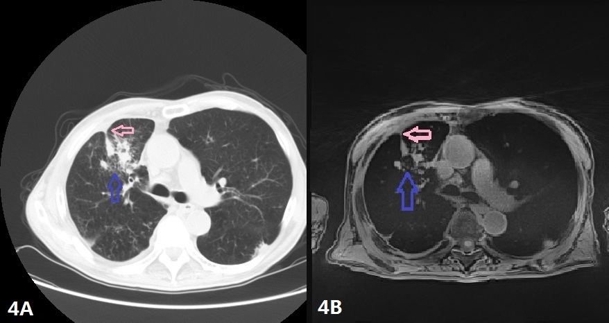
Figure 4A-4B A 43-year–old male with patchy shadow(Blue arrow) and pleural thickenings(Pink arrow),these signs can be displayed clearly CT (Figure 4A)as well as on Star VIBE(Figure 4B).
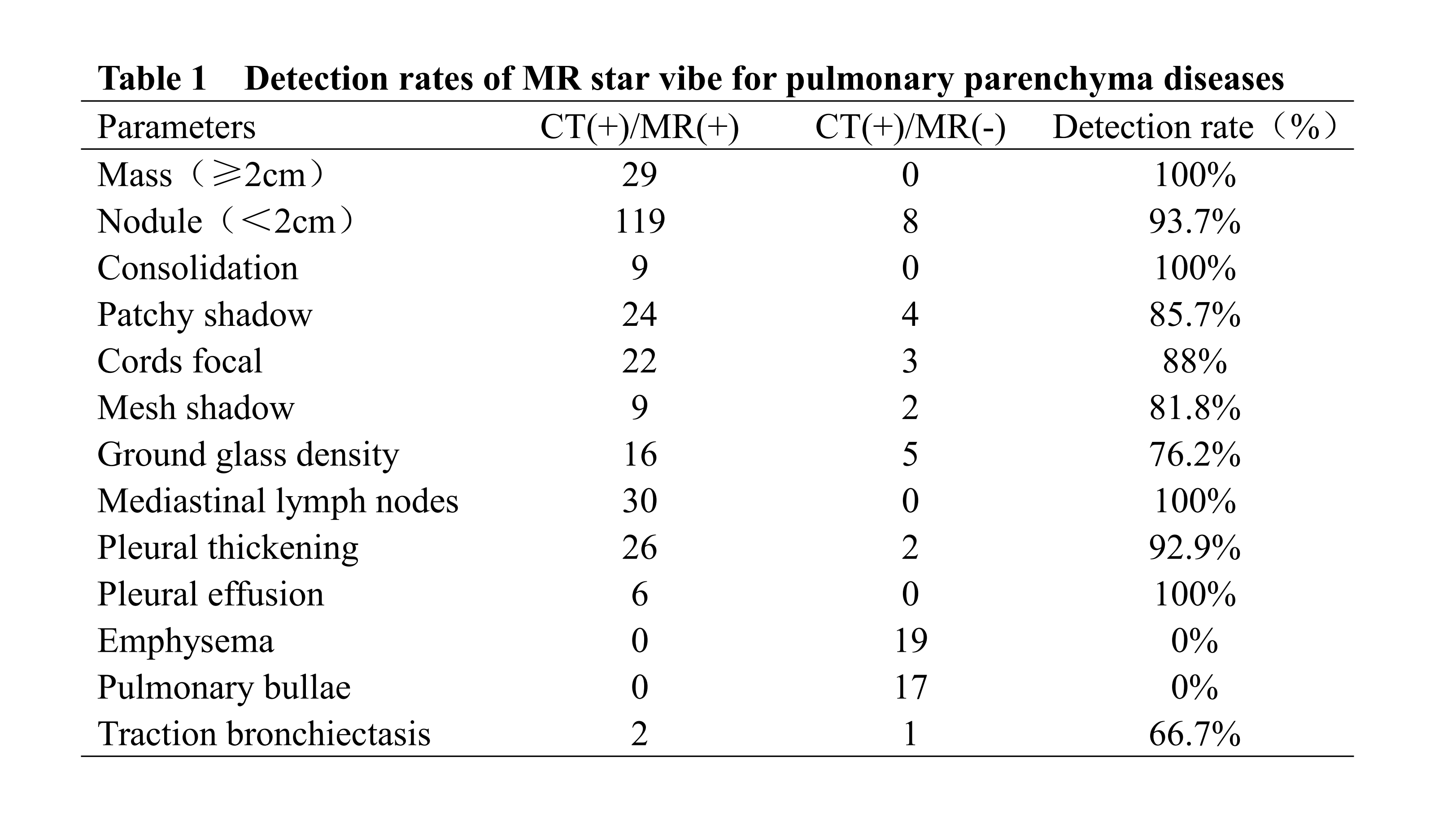
Table1