2440
Fast Imaging of Hyperpolarized Xe-129 in the Airspace, Barrier and Red Blood Cells in the Human LungJunshuai Xie1,2, Haidong Li1, Huiting Zhang1, Xiuchao Zhao1, Xianping Sun1,2, Chaohui Ye1,2, and Xin Zhou1,2
1State Key Laboratory of Magnetic Resonance and Atomic and Molecular Physics, National Center for Magnetic Resonance in Wuhan,Wuhan Institute of Physics and Mathematics, Chinese Academy of Sciences, Wuhan, China, 2University of Chinese Academy of Sciences, Beijing, China
Synopsis
Xe-129 in the barrier and red blood cells could be separated by the dissolved-phase (DP) Xe-129 MRI with radial sampling strategy. However, the number of the RF pulse was usually large and thus resulted in long acquisition time. An MRI strategy in the Cartesian coordinate has been used to for high-resolution rodent lung imaging of He-3 in the airspace. The concept was introduced into fast acquisition of the DP Xe-129 in the human lung with the multi-point Dixon method. The number of the RF pulse reduced and the results of TP/Gas and RBC/Gas agreed with the previous study.
Introduction
Xe-129 in the barrier (TP) and red blood cells (RBC) could be separated by the dissolved-phase (DP) Xe-129 MRI with radial sampling strategy and Dixon methods1,2. However, the number of the RF pulse was usually large and thus resulted in long acquisition time. An MRI strategy in the Cartesian coordinate has been used to for high-resolution rodent lung imaging of He-3 in the airspace (Gas)3, which could reduce the number of the RF pulse compared to the radial sampling strategy. The concept was introduced into fast acquisition of the DP Xe-129 in the human lung with the multi-point Dixon method.Methods
The pulse sequence developed in this study was shown in Fig. 1. An optimized 3D FLASH sequence (BAMI) was used for data acquisition with compressed sensing1. For each of the phase-encode step, the multi-echo data of the DP and Gas signals of Xe-129 were both acquired. A 1.2 ms sinc RF pulse (centered at +217 ppm relative to the resonance frequency of xenon in airspace) was used to excite the dissolved-phase xenon. And proton images were obtained in the same breath-hold with conventional 3D FLASH sequence. All the experiments were performed on a 1.5-T commercial whole-body imager (Avanto, Siemens Medical Solutions, Malvern, PA) using a homemade transmit-receive vest RF coil. Enriched xenon (86% 129-isotope) was polarized using a homemade xenon polarizer. One liter of xenon mixture (70% xenon + 30% N2) was inhaled from functional residual capacity before each measurement. Informed consent was obtained from each of 4 healthy young subjects (aged between 23 and 27, HY). The protocol was approved by the Ethics Committee of the Wuhan Institute of Physics and Mathematics, Chinese Academy of Sciences (WIPM, CAS). The multi-point Dixon method was used to separate the TP and RBC signals. And the ratios of DP/Gas, TP/Gas and RBC/Gas were calculated.Results
The ratio maps of TP/Gas and RBC/Gas were presented in Fig. 2 with the relevant proton images. For the HY subjects, DP/Gas = 1.36% ± 0.20%, TP/Gas = 0.96% ± 0.16% and RBC/Gas = 0.40% ± 0.07%. These results agreed with the previous study1.Conclusion
Xe-129 in the airspace, barrier and red blood cells in the human lung were successfully obtained in a single breath-hold via hyperpolarized Xe-129 MRI. The method BAMI could reduce the number of the RF pulse compared to the radial sampling strategy, which is useful in the clinical practice.Acknowledgements
We acknowledge the support by the Natural Science Foundation of China (81227902, 81625011, 81601491, 81771917) and National Program for Support of Eminent Professionals (National Program for Support of Top-notch Young Professionals).References
1. Kun Qing et al. NMR Biomed 2014; 27: 1490-1501.
2. Kaushik SS et al. Magn Reson Med 2016; 75: 1434-1443.
3. Ouriadov AV et al. ISMRM 2008: p3789.
Figures
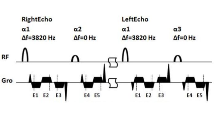
Figure 1.The
pulse sequence diagram of BAMI. For each of the phase-encode step, the multi-echo
data of the dissolved-phase (with echo time 1.00 ms, 2.26 ms and 2.52 ms) and
gas-phase(with echo time 1.00 ms and 2.26 ms) signals of Xe-129 were both
acquired. A 1.2 ms sinc RF pulse (centered at +217 ppm relative to the resonance
frequency of xenon in airspace) was used to excite the dissolved-phase xenon. The
flip angle α1
was 35° and α2 0.5° and α2 = asin(tanα2).
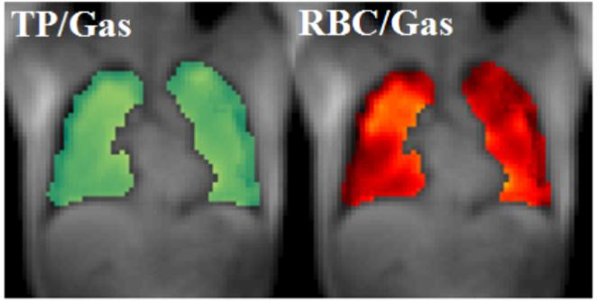
Figure 2. The ratio maps of TP/Gas and RBC/Gas in one healthy
young subject were presented with the relevant proton images. For
the healthy
young subjects, DP/Gas = 1.36% ± 0.20%,
TP/Gas = 0.96%
± 0.16% and RBC/Gas = 0.40% ± 0.07%.
These results agreed with the previous study1.