2439
Using a hybrid multibreath hyperpolarized (HP) 129Xe imaging technique for simultaneous assessment of lung function and structure in a two-hit radiation induced lung injury (RILI) model.1Radiology, University of Pennsylvania, Philadelphia, PA, United States, 2Physiology, University of Pennsylvania, Philadelphia, PA, United States
Synopsis
In this study we developed a two-hit hemi-thorax radiation-induced lung injury (RILI) model that better simulates the etiology of the disease in humans, and characterized it via a multibreath hyperpolarized (HP) 129Xe imaging technique to assess lung function and structure one month post-radiation. We observed an increased PAO2 of 145±41 Torr in the radiated lung compared to 124±40 Torr in the contralateral lung. We also observed a corresponding decrease in oxygen uptake in the radiated lung. The preliminary findings suggest that HP 129Xe-derived functional parameters, particularly changes in the alveolar oxygen tension and oxygen uptake can serve as biomarkers during the early fibrotic stage of RILI.
Introduction
Radiation
induced lung injury (RILI) is a major side effect in patients
undergoing thoracic irradiation for lung or breast cancer [1]. The
injury typically manifests as pneumonitis before progressing to
irreversible pulmonary fibrosis. Unfortunately, the conventional rat
models are not ideal for studying this phenomenon, due to their
relative insensitivity to RILI at dosages comparable to those used
for radiation therapy (RT) in humans [2]. In this study, we developed
a two-hit RILI model that better simulates the etiology of the
disease in humans, and characterized it via a multibreath
hyperpolarized (HP) 129Xe imaging technique to assess lung
function and structure one month post-radiation.Methods
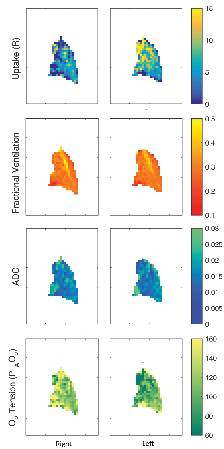
The two-hit RILI model consists of priming the lungs with an intratracheal instillation of LPS (10mg/mL) followed by right hemi-thorax radiation (25Gy) after 24 hours, in Fisher rats (n=8). Animals were imaged one month post-radiation, when early fibrosis can typically be detected. Gated micro-computed tomography (uCT) (current = 60mA, voltage = 40kV, reconstructed isotropically at 200 um) was used to detect structural changes. A 1.5T MRI system (Seimens MAGNETOM) with a custom-built quadrature volume coil was used to to perform a hybrid multibreath hyperpolarized 129Xe imaging sequence that was used to derive the following functional parameters: fractional ventilation (FV), alveolar oxygen tension (PAO2), apparent diffusion coefficient (ADC), and oxygen uptake (R) [3]. The fractional ventilation image series consisted of 10 wash-in breaths followed by a final long breath-hold for T1 and flip angle correction, as previously presented [3]. Two 10mm sagittal slices with a 2D multi-slice GRE sequence were acquired with a matrix size of 48x36 (3mm x 3mm in-plane resolution), flip-angle = 6o, and TR/TE=9.0/4.1ms.Results and Discussion
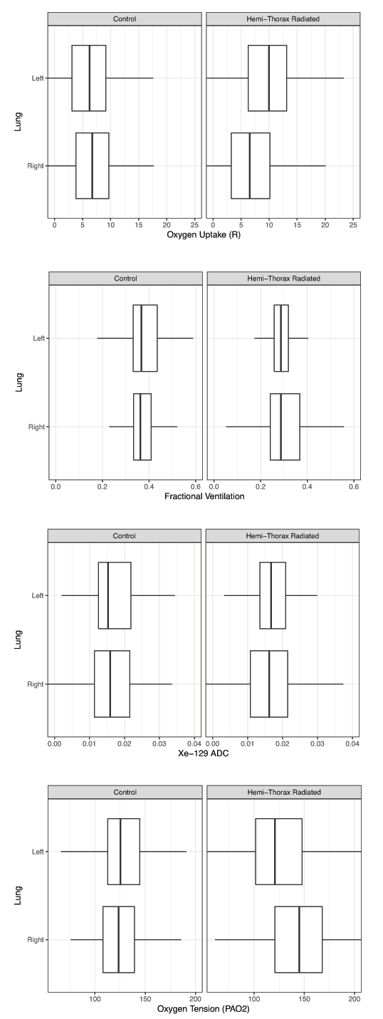
The preliminary findings show
that the PAO2 in the radiated lungs
significantly increased to 145±41 Torr, compared to 124±40 Torr in
the contralateral lungs, and 125±39 Torr in the healthy cohort (both
lungs). On the other hand, the radiated lungs had an R of 6.3±6.1
Torr/s, compared to 9.8±7.3 Torr/s in the contralateral lungs. The
control cohort had a R of 7.0±10.3 Torr/s, which suggests that the
lowered oxygen uptake in the radiated lungs may have been compensated
by an increased uptake in the contralateral lung. Physiologically,
these observed changes in R are most likely due to the increased
alveolar wall thickness with the onset of fibrosis. Although no
significant differences were observed for FV and ADC between the
radiated lungs and the other cohorts, the heterogeneity in their
measurements may serve as a potential biomarker as well.Conclusions
Conclusions:
This study demonstrates that HP 129Xe-derived functional
parameters, particularly changes in the alveolar oxygen tension and
oxygen uptake can serve as biomarkers during the early fibrotic stage
of RILI. The next goal of this study is to quantify these changes at
earlier timepoints (pneumonitis stage) of the disease in order to
determine the sensitivity of the imaging technique and its
feasibility for early detection of RILI.Acknowledgements
No acknowledgement found.References
[1] Graham, Intl J Radiat Oncol Biol Physics, 1999:45
(2):323 [2] Haston, Cancer Res, 2007:67(22):10796-803
[3] Hamedani, MRM, 2017:78(2):611-624
Figures