2437
Revealing Pulmonary Gas Transport Dynamics using a 3D Radial Hyperpolarized Xenon MRI Acquisition with Variable Flip Angles1Radiology, University of Pennsylvania, Philadelphia, PA, United States
Synopsis
We demonstrated that reducing the flip angle drives the distribution of acquired dissolved-phase xenon downstream towards the heart. By exploiting this principle, the dynamics of pulmonary gas transport were captured through a single 3D double golden means radial acquisition with linearly decreasing flip angles. Reconstruction with a sliding window generated a series of consecutive images with declining average flip angles, depicting the gradual uptake and accumulation of xenon by the heart and lungs.
Introduction
Hyperpolarized xenon-129 (HXe) is a powerful agent for evaluating pulmonary gas uptake and transport given its ability to diffuse from alveoli into the blood and flow into the heart. Existing imaging techniques typically capture a snapshot of xenon dissolved in the lung parenchyma, with the depicted distribution determined by the combination of measurement repetition time and flip angle1-6. In this study, we investigated the feasibility of obtaining information about xenon transport dynamics in a single breath-hold by linearly varying the flip angle throughout the measurement using a 3D double golden means radial acquisition7.Methods
Imaging experiments were performed in 2 sedated New Zealand rabbits (approx. 4.5 kg), and were approved by the Institutional Animal Care and Use Committee. Animals were ventilated with room air until imaging began, when the gas mix was switched to 20% oxygen and 80% HXe for two breaths (6 ml/kg tidal volume); acquisition began on the second breath. Images were acquired using a 3D double golden means radial sequence with interleaved gas- and dissolved-phase excitations. Parameters included TR/TE = 8.32/0.62 ms, 1,600 radial spokes with 128 sampling points, dwell time of 5 ms, and FOV of 180-300 mm3. The flip angle was either held fixed at 2° or 20°, or decreased linearly from 10° to 1° over the course of the acquisition. Radial data sets with 1,600 and 400 spokes were gridded onto 32x32x32 and 22x22x22 matrices, respectively, and Fourier transformed using the Michigan Image Reconstruction Toolbox. The double golden means sampling ensured relatively uniform k-space sampling regardless of the number of collected spokes. A window of 400 and step size of 10 spokes were used to reconstruct the variable flip angle acquisition in a sliding-window fashion, such that the average flip angle for each subsequent reconstructed 3D data set decreased slightly from the one reconstructed before. All dissolved-phase images were normalized to their respective gas-phase images. The sliding window data sets from the variable flip angle acquisition were normalized to the reconstructed dissolved-phase image with the highest average flip angle.
All MR studies were performed at 1.5T (Avanto; Siemens), using a custom xenon-129 transmit/receive birdcage coil (Stark Contrast, Erlangen, Germany). Enriched xenon gas (87% xenon-129) was polarized using a prototype commercial system (XeBox-E10, Xemed LLC, Durham, NH).
Results and Discussion
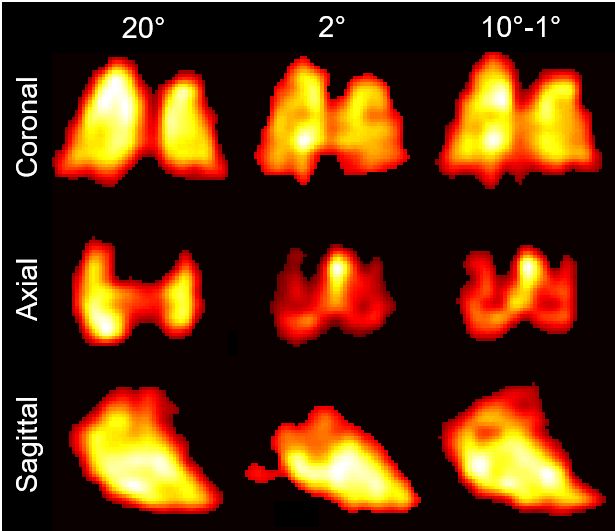
The effect of different flip angles can be seen in Fig. 1, which shows the same dissolved-phase slices with acquisition flip angles of 20°, 2°, and varying linearly from 10° to 1°. Reducing the flip angle from 20° to 2° shifted the depicted dissolved-phase distribution downstream, away from the alveolar exchange sites and into the heart. This is particularly evident in the axial view, where the xenon is predominantly present in the lungs at 20°, while at 2° the xenon is consolidated in the heart. The varying flip angle acquisition straddles these extremes by exhibiting strong dissolved-phase signals both upstream in the lung parenchyma and further downstream in the heart. This observed flip angle dependence is the result of the xenon transported further from the alveolar exchange sites experiencing more RF pulses. Hence, in measurements with higher flip angles, downstream xenon magnetization is quickly depolarized and is therefore invisible in the reconstructed images.
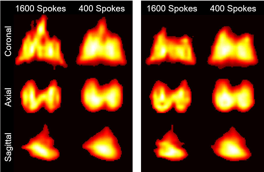
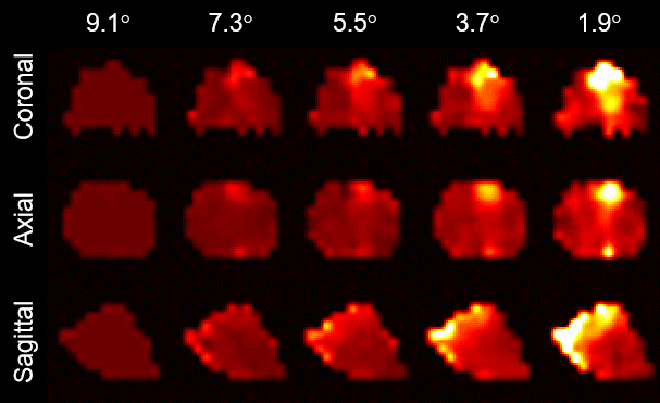
Figure 2 illustrates the resulting loss in image resolution when only 400 spokes instead of the sampled 1,600 spokes are used for image reconstruction. However, by reconstructing only 400 spokes of the variable flip angle acquisition at a time and then continually shifting the reconstruction window by 10 spokes, each calculated 3D image set will be based on a measurement with a slightly lower average flip angle than the one before. As demonstrated in Fig. 3, the resulting image series reveals a detailed, gradual visualization of the xenon gas uptake dynamics in the parenchyma and subsequent accumulation in the left side of the rabbit heart.
Conclusion
We demonstrated that a single 3D double golden means acquisition can capture the gradual uptake and transport of xenon gas by the pulmonary system by employing linearly decreasing flip angles.Acknowledgements
Supported by NIH grants R01 EB015767 and R01 HL129805.References
[1] Driehuys et al. Imaging alveolar–capillary gas transfer using hyperpolarized 129Xe MRI. Proc Natl Acad Sci USA 2006; 103:18278-18283.
[2] Patz et al. Human Pulmonary Imaging and Spectroscopy with Hyperpolarized 129Xe at 0.2T. Acad Radiol 2008; 15:713-727.
[3] Mugler et al. Simultaneous magnetic resonance imaging of ventilation distribution and gas uptake in the human lung using hyperpolarized xenon-129. Proc Natl Acad Sci USA 2010;107(50):21707-21712.
[4] Cleveland et al. Hyperpolarized 129Xe MR Imaging of Alveolar Gas Uptake in Humans. Plos One 2010; 5:e12192.
[5] Qing et al. Regional Mapping of gas uptake by blood and tissue in the human lung using hyperpolarized xenon-129 MRI. J Magn Reson Imaging 2013; 39(2):346-359.
[6] Doganay et al. Quantification of regional early stage gas exchange changes using hyperpolarized 129Xe MRI in a rat model of radiation-induced lung injury. Magn Reson Med 2016; 76:566-576.
[7] Chan et al. Temporal stability of adaptive 3D radial MRI using multidimensional golden means. Magn Reson Med 2009; 61(2):354-363.
Figures