2433
Regional Lung Function Quantification by Combining Gas-Phase Saturation with Hyperpolarized Xenon-129 Dissolved-Phase MRI1Radiology, University of Pennsylvania, Philadelphia, PA, United States
Synopsis
Hyperpolarized xenon-129 MRI has previously been used to assess pulmonary gas exchange between the alveolar volume and lung tissue. In this work, we quantified changes in the downstream xenon dissolved-phase signal in the left ventricle in response to a regional saturation of the pulmonary gas-phase signal. This approach permitted us to extract the relative gas-exchange efficiency of the lung volume unaffected by the GP signal saturation, demonstrating increased gas exchange efficiency in the posterior regions of the lung in supine rabbits. The proposed technique might be especially valuable in lung transplantation, during pharmaceutical interventions, or for lung-volume reduction surgeries.
Purpose
Existing imaging techniques for detecting lung disorders are unable to measure the degree to which functional impairment of a specific region of the lung contributes to an overall reduction in gas transport. Hyperpolarized xenon-129 (HXe) MRI has previously been used to assess pulmonary gas exchange between the alveolar volume and lung tissue1-7. In this work, we quantified changes in the downstream HXe dissolved-phase (DP) signal in the left ventricle in response to a regional saturation of the pulmonary gas-phase (GP) signal. This approach permitted us to extract the relative gas-exchange efficiency of the lung volume unaffected by the GP signal saturation.Methods
Imaging experiments were performed in two sedated New Zealand rabbits (approx. 4.5 kg). Animals were ventilated with room air until imaging began, at which point the gas mix was switched to 20% oxygen and 80% HXe for two breaths (6 ml/kg tidal volume). Imaging was performed at end inspiration of the second breath. All studies were approved by the Institutional Animal Care and Use Committee. MR imaging was conducted using a 2D-projection gradient-echo sequence that employed a non-selective 900-μs Gaussian RF excitation pulse centered 3,530 Hz downfield from the GP resonance. The large frequency difference between the two phases, combined with a sufficiently small acquisition bandwidth, allowed us to simultaneously image HXe in the pulmonary air spaces and dissolved in the lung tissue side-by-side4. The following sequence parameters were used: matrix size 36×80; TR 200 ms; TE 2.7 ms; flip angle 40° or 75°; FOV 238 mm; receiver bandwidth 110 Hz/pixel. Immediately preceding the data acquisition, a regional RF saturation pulse was applied to destroy the HXe GP magnetization in the selected lung volume. To quantify regional lung function, the average HXe signal in the left ventricle was calculated following saturation of the GP signal in a coronal slab and normalized by the residual GP signal. The saturation slab was moved from anterior to posterior in 5 mm increments, thereby increasing the saturation volume in a step-wise fashion. All MR studies were performed at 1.5T (Avanto; Siemens), using a custom xenon-129 transmit/receive birdcage coil (Stark Contrast, Erlangen, Germany). Enriched xenon gas (87% xenon-129) was polarized using a prototype commercial system (XeBox-E10, Xemed LLC, Durham, NH).Results and Discussion
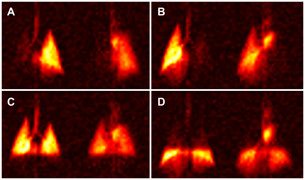
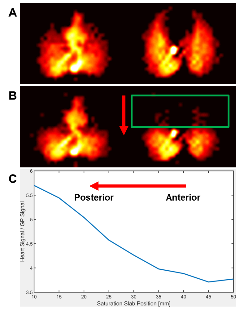
The coronal 2D projection images in Figure 1 demonstrate the impact of selectively saturating the GP in various lung regions. This process also depletes the DP magnetization in those volumes of the lung with which the saturated GP region stands is exchanging. The remaining DP signal originates from gas exchange in, and subsequent transport from, the lung volumes unaffected by the GP saturation.Figure 2A shows axial projections of simultaneously-acquired DP (left) and GP (right) images in a healthy rabbit. Note that in the axial orientation GP and DP positions in each image panel are swapped with respect to Fig. 1. Figure 2B depicts the same acquisition as Fig. 2A, but following a regional saturation pulse that destroyed the GP signal in the selected coronal slab. As the saturation slab is moved from the anterior towards the posterior of the lung, the normalized HXe signal in the left ventricle increases (Fig. 2C), which corresponds to increased gas exchange efficiency in the posterior regions of the lung in supine rabbits. It is worth pointing out that all our measurements for this study were conducted at end inspiration, but that we anticipate that the regional gas exchange efficiency may vary throughout the respiratory cycle.Conclusion
In this work, we demonstrated how HXe MRI can be used to quantify the contributions of specific lung volumes to overall lung function. Specific lung geometries can be assessed by applying appropriately-designed saturation pulses; this might be especially valuable in lung transplantation, during pharmaceutical interventions, or in the planning of lung-volume reduction surgeries.Acknowledgements
Supported by NIH grants R01 EB015767 and R01 HL129805.References
[1] Ruppert et al. NMR of hyperpolarized 129Xe in the canine chest: spectral dynamics during a breath-hold. NMR Biomed 2000;13:220-228.
[2] Driehuys et al. Imaging alveolar–capillary gas transfer using hyperpolarized 129Xe MRI. Proc Natl Acad Sci USA 2006;103:18278-18283.
[3] Patz et al. Human Pulmonary Imaging and Spectroscopy with Hyperpolarized 129Xe at 0.2T. Acad Radiol 2008;15:713-727.
[4] Mugler et al. Simultaneous magnetic resonance imaging of ventilation distribution and gas uptake in the human lung using hyperpolarized xenon-129. Proc Natl Acad Sci USA 2010;107(50):21707-21712.
[5] Cleveland et al. Hyperpolarized 129Xe MR Imaging of Alveolar Gas Uptake in Humans. Plos One 2010;5:e12192.
[6] Qing et al. Regional Mapping of gas uptake by blood and tissue in the human lung using hyperpolarized xenon-129 MRI. J Magn Reson Imaging 2013; 39(2):346-359.
[7] Doganay et al. Quantification of regional early stage gas exchange changes using hyperpolarized 129Xe MRI in a rat model of radiation-induced lung injury. Magn Reson Med 2016;76:566-576.
Figures