2425
Diffusion tensor and Intravoxel incoherent motion magnetic resonance imaging of the normal breast in young premenopausal women during menstrual cycleQiuju Fan1, Hui Tan1, Nan Yu1, Qi Yang1, Shaoyu Wang2, and Yong Yu1
1Affiliated Hospital of Shaanxi University of Chinese Medicine, Xianyang, China, 2Siemens Healthcare, Scientific marketing, Shanghai, China
Synopsis
DTI and IVIM can provide valuable information on tissue microstructure, microcirculation and pathophysiology that has been extensively used on the breast cancer [1,2].However, the breast is a hormonally responsive organ and undergoes periodic variations according to the menstrual cycle. Thus, the periodic variations of DTI and IVIM-derived measurements need to be considered.
Introduction
Our goal was to investigate the parameters obtained with DTI and IVIM imaging of the breast throughout the menstrual cycle phases in volunteers.Materials and method
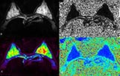
The study included 26 healthy female volunteers (median age, 26 years; age range, 22–30 years), with a regular menstrual cycle, none of the volunteers were taking oral contraceptives, and all were nulliparous. These volunteers underwent imaging five times during one menstrual cycle: (menstrual, proliferative, follicular, luteal and secretory). All data were collected on 3T MR scanner (MAGNETOM Skyra, Siemens Healthcare, Erlangen, Germany), a receiving four-channel breast array coil. The imaging protocols included axial T2-weighted, DTI and IVIM sequences. Axial DTI images with fat suppression and single-shot EPI imaging sequence, with diffusion gradients in 30 directions during 10 minutes, b values of 0 and 700 sec/mm2. Then IVIM sequences using single-shot EPI sequence, fat suppression techniques, b values of 0, 50, 100, 150, 200,250, 300, 400, 600, 800 sec/mm2, each b-value was acquired for each of three orthogonal directions.Results
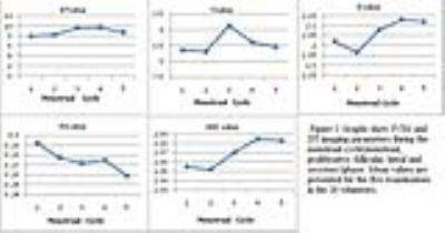
Quantitative parameters including perfusion fraction (f), molecular diffusion coefficient (D), and microperfusion coefficient (D*) were used in IVIM to evaluate the molecular diffusion of water molecules in the tissues and microvascular perfusion. DTI related parameters is fractional anisotropy (FA) and ADC reflects. The values were analyzed of Univariate Analysis of Variance. For normal breast, the D* values showed regular fluctuations: D* values is higher in luteal phase than in other phase, there were significant differences during menstrual and proliferative phases compare to luteal phase(p<0.01); In the follicular phase, f values are higher than in other phases, but, no significant differences were found. The standard deviation of f values is particularly difference. Similar results were found for D and ADC values. D values have shown proliferation increases in the luteal vs. proliferative, the difference was statistically significant (p = 0.005). During menstrual cycle, the difference of ADC did not reach statistical significance in this study. FA value in the menstrual period compared to the secretory phase meaningful (P=0.034).Discussion and Conclusions
MRI is sensitive to hormone-induced breast tissue changes during the menstrual cycle. The IVIM parameters D* values determined in our study mainly reflected the vascularity of the breast tissue fluctuates in response to hormonal changes. The diffusion coefficients and the anisotropy indexes were not very sensitive to the edema in interlobular stroma that occurred during the menstrual cycle.Acknowledgements
No acknowledgement found.References
1. Baltzer PA, Schafer A, Dietzel M, et al.Diffusion tensor magnetic resonance imaging of the breast: a pilot study. Eur Radiol 2011;21(1):1–10.
2. Eyal E, Shapiro-Feinberg M, Furman-HaranE, et al. Parametric diffusion tensor imagingof the breast. Invest Radiol 2012;47(5):284–291.

