2424
Proton MR Spectroscopy in Breast: Lipid Metabolite Concentrations as Valuable Quantitative Imaging Biomarkers for Cancer Diagnosis1Medical Physics, Memorial Sloan Kettering Cancer Center, New York, NY, United States, 2Radiology, Memorial Sloan Kettering Cancer Center, New York, NY, United States, 3GE Global Research, Niskayuna, NY, United States, 4Memorial Sloan Kettering Cancer Center, New York, NY, United States, 5Medical University Vienna, Vienna, Austria
Synopsis
Differential expression of lipid metabolism-related proteins was recently
reported in breast cancer patients. In this retrospective MR spectroscopy (MRS)
study, the spectral lipid profile was assessed in breast cancer patients with
malignant and benign lesions. Single-voxel MRS data from 176 breast lesions was
analyzed to quantify multiple lipid metabolite concentrations using LCModel. Lipid peak analysis
highlighted significant differences in lipid
metabolite concentrations with significantly low concentrations in
malignant compared to benign lesions and in luminal cancers compared to other molecular
subtypes. MRS-based lipid metabolite profile
may provide a valuable tool for breast cancer diagnosis.
Introduction
In breast cancer diagnosis, the value of proton MR spectroscopy (1H-MRS) has been largely based on the detection of choline (Cho) metabolites for the differentiation of benign and malignant tumors or the spatio-longitudinal monitoring of changes in Cho metabolites during neoadjuvant chemotherapy1. However, in addition to the Cho metabolites, multiple other metabolites including lipids can be detected and monitored with 1H-MRS2-4. Initial ex vivo NMR and in-vivo MRS studies have shown differences in fat metabolites and water between malignant, benign and normal tissue with higher water concentrations and lower methylene lipid peaks at 1.3 ppm in breast cancer. Although research so far has focused on the detection of the 1.3 ppm lipid peak, a whole spectrum of lipid peaks can be detected with MRS. We hypothesize that through the non-invasive quantitative assessment of lipid metabolites with 1H-MRS insights into tumor biology are feasible, which might be useful for the breast cancer diagnosis and differentiating molecular subtypes.Purpose
The aim of this study was to investigate whether lipid metabolite concentrations detected with 1H-MRS can be used for a non-invasive differentiation of (1) benign and malignant breast tumors and (2) luminal and non-luminal molecular breast cancer subtypes.Materials and Methods
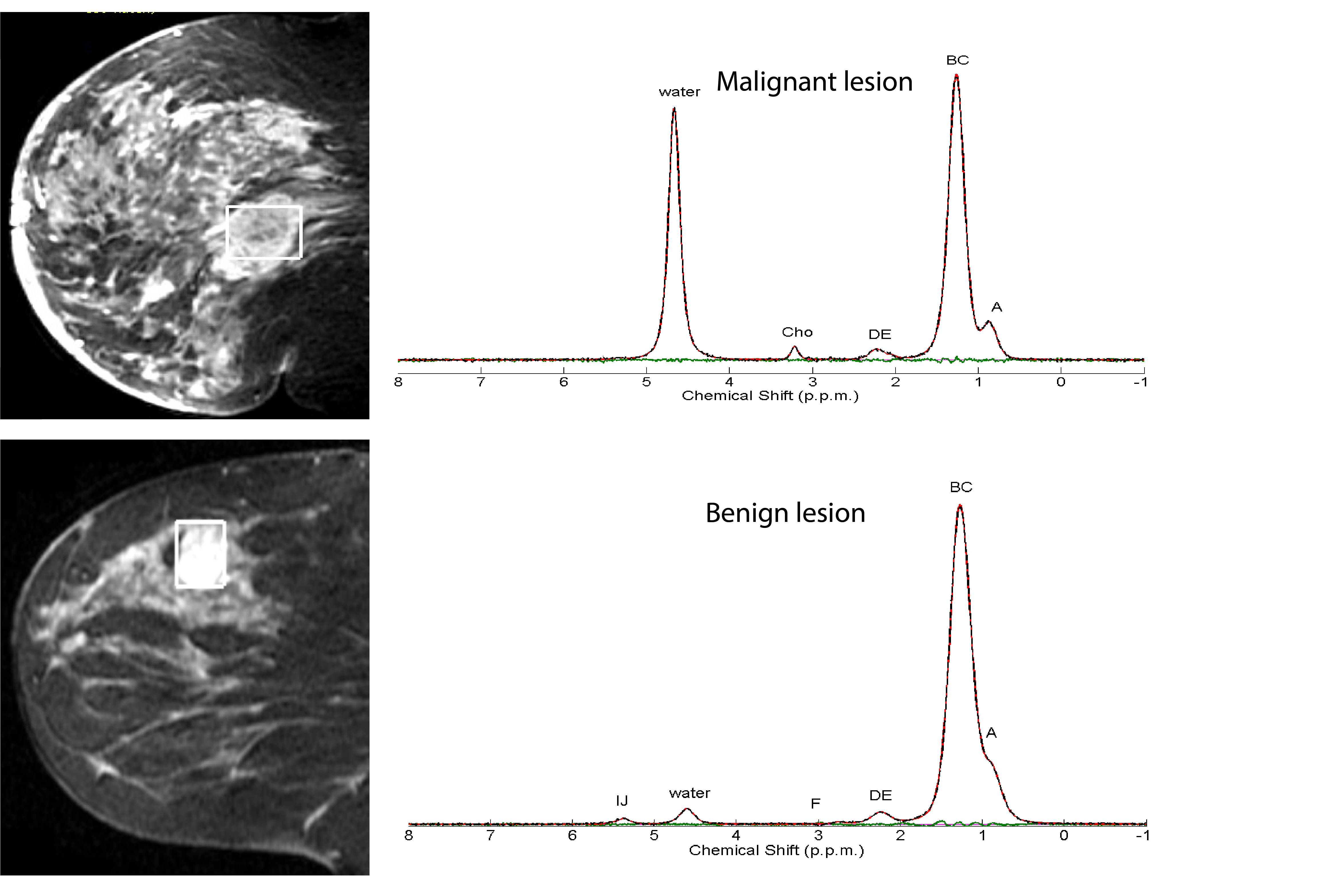
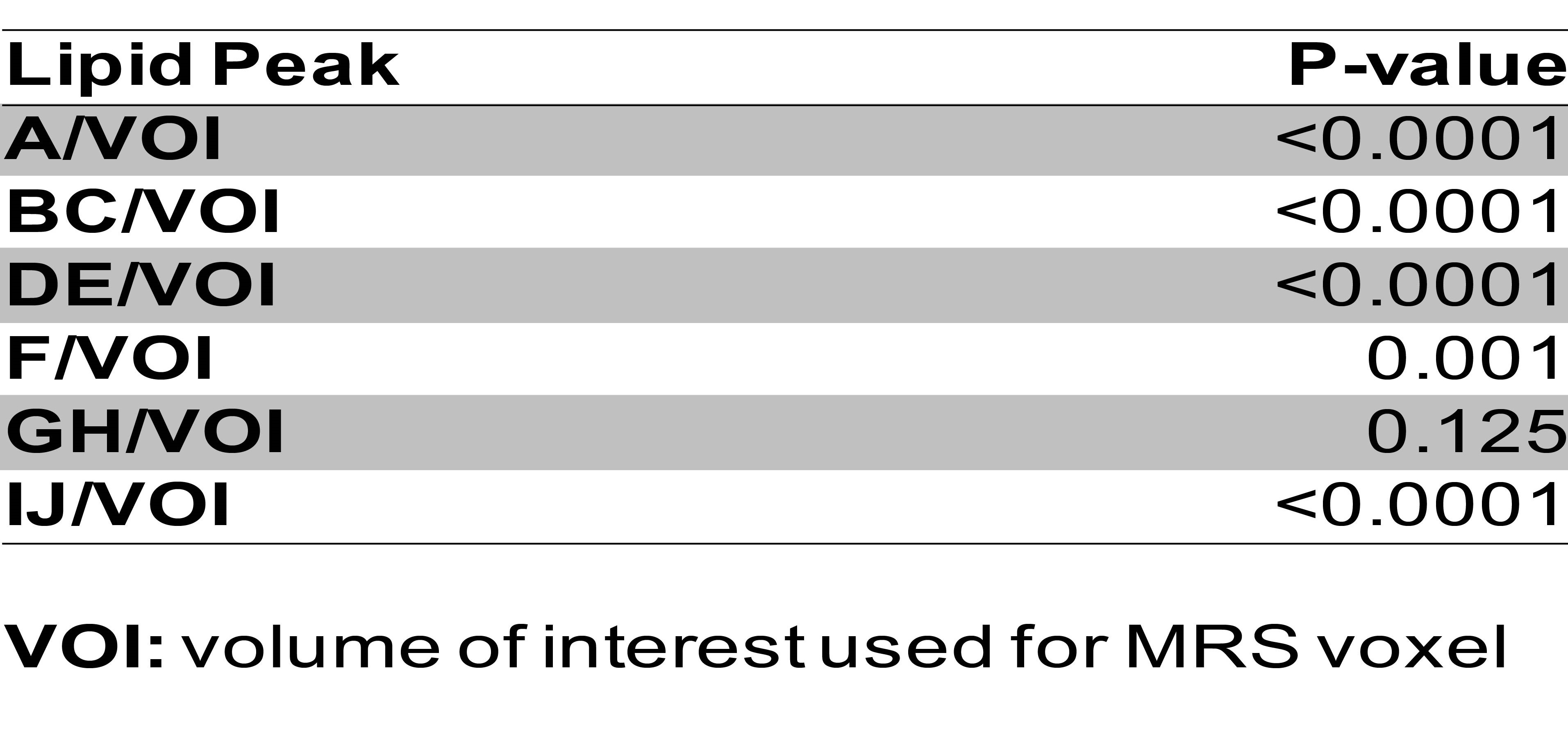
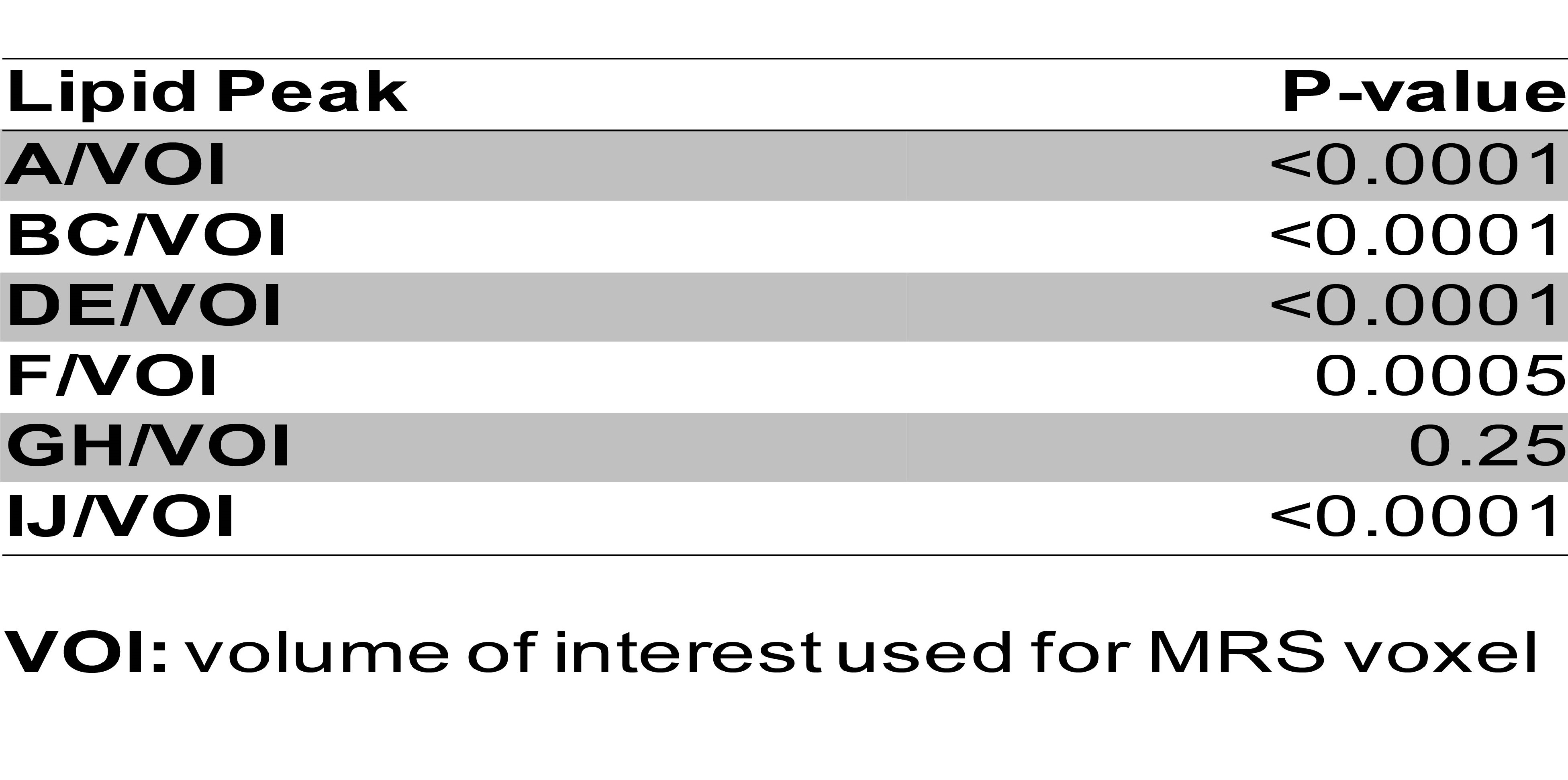
In this HIPPA-compliant, IRB-approved study, data of 176 women who underwent MRI with T2 weighted, dynamic contrast-enhanced MRI and 1H-MRS at 1.5T for either a suspicious imaging finding or staging of a known cancer (BI-RADS 0,4/5/6) were retrospectively analyzed. In all patients single-voxel 1H-MRS data were collected using a PRESS MRS Sequence with TR/TE=2000/135ms. Quantitative analysis of all lipid resonances present in the 1H-MRS data was done using LCModel analysis. Metabolite CRB values less than 20% were excluded from analysis as non-reliable measurements. We classified all metabolites into 6 groups based methyl, methylene, and methine proton chemical shifts, such as A (0.9ppm), BC (1.3 and 1.59 ppm), DE (2.04 and 2.25 ppm), F (2.77 ppm), GH (4.1 and 4.25 ppm), IJ (5.22 and 5.31 ppm). Lipid concentrations were normalized with MRS volume of interest (VOI). Bivariate and ROC Curve analyses were performed to determine associations, and the predictive accuracy of 1H-MRS imaging biomarkers with pathological tumor characteristics (benign vs. malignant, molecular subtypes [Luminal A/B versus others: Her2-enriched and Triple negative), tumor grade, nodal status, lymphovascular invasion (LVI)]. Histopathology was used as the standard of reference.Results
There were 176 lesions including 85 invasive ductal cancers, 10 invasive lobular cancers, 13 ductal carcinoma in situ, 6 other types of breast cancer, and 62 benign lesions. The mean and std dev of voxel size was 4.4 ± 4.6 cm. All 1H-MRS lipid resonances were successfully quantified (Figure 1). A, BC, DE, F and IJ lipid metabolite concentrations were significantly lower in malignant compared to benign tumors (Table 1) as well as in luminal compared to other subtypes (Table 2) (p < 0.001). There was no significant difference in lipid metabolite concentrations in malignant lesion based on histological grade, LIV and axillary lymph node metastases.Conclusion
Lipid metabolite concentrations quantified with 1H-MRS can successfully detect breast cancer and differentiate luminal breast cancers from other more aggressive breast cancer subtypes. Lipid metabolite concentrations quantified with 1H-MRS may therefore provide a valuable imaging biomarker in addition to choline.Acknowledgements
NoneReferences
1. Bolan et al. Magnetic resonance spectroscopy of the breast: current status. Magn Reson Imaging Clin N Am. 2013
2. Thakur S et al., Comparisons of Water-to-Fat Ratios in Malignant, Benign Breast Lesions, and Normal Breast Parenchyma: An In Vivo Proton MRS Study. ISMRM 2006
3. Thakur S et al. Discrimination of Choline-Positive Invasive Breast Carcinomas Using Water-to-Fat Ratio: A Proton MRS Study. ISMRM 2006
4. Freed et al. Evaluation of Breast Lipid Composition in Patients with Benign Tissue and Cancer by Using Multiple Gradient-Echo MR Imaging. Radiology. 2016
Figures