2409
Improving the resting state fMRI detection in anesthetized monkeys using multiband MRI techniqueChun-Xia Li1, Doty Kempf1, Leonard Howell1,2, and Xiaodong Zhang1,2
1Yerkes Imaging Center, Yerkes National Primate Research Center, Emory University, Atlanta, GA, United States, 2Division of Neuropharmacology and Neurologic Diseases, Yerkes National Primate Research Center, Emory University, Atlanta, GA, United States
Synopsis
Neuroimaging
studies of non-human primates are generally conducted with anesthesia using anesthetics
like isoflurane which is known to suppress the neuronal activation of the brain
substantially. The resting state functional MRI (rsfMRI) examination in
anesthetized monkeys is hindered by limited choices of anesthetics compared to
rodent studies. In the present study, multiband MRI technique was explored to
improve the rsfMRI detection in anesthetized macaque monkeys. The preliminary
results suggest the multiband MRI can be employed to dramatically improve the
rsfMRI detection in examining the functional connectivity of default mode
network in anesthetized monkeys using a clinical 3T setting.
Introduction
Resting-state functional MRI (rsfMRI) has become
a robust approach widely used to examine the resting state brain networks in
preclinical and clinical studies. However, most preclinical neuroimaging
studies are performed on sedated animals with anesthetics like isoflurane which
is known to suppress the neuronal activation of the brain substantially. In
order to improve the rsfMRI signal detection in anesthetized animals, ultra-high
field MRI is applied together with optimal anesthesia procedures to minimize
the neuronal suppression effects, and combined usage of medetomidine and
isoflurane is suggested to be an optimal option for rsfMRI in rodent models [1].
However, medetomidine is not recommended for routine usage in monkeys due to
its side effects in non-human primates (NHPs). Ultra-high field MRI scanners
are usually not accessible for most neuroimaging studies with large animals. rsfMRI studies in NHPs are still hindered by
the usage of popular anesthetics like isoflurane and ketamine. Previous studies have demonstrated that multiband
MRI technique can improve the rsfMRI detection sensitivity and statistical
power substantially in studies of human connectome [2]. In the present study, the
multiband MRI technique was explored for rsfMRI examination in anesthetized macaque
monkeys using a clinical setting.Methods
Adult rhesus monkeys (7-11 years old, 8-10kg) were anesthetized with 1% isoflurane mixed with 100% oxygen. Physiological parameters such as O2 saturation, blood pressure, heart rate, respiration rate, body temperature and PaCO2 were monitored continuously and maintained in normal ranges [3]. rsfMRI data were acquired using the multiband EPI sequence (TR=0.590ms, 1000s, 2000s, TE=25ms, FOV = 96 mm × 96 mm, spatial resolution= 1.5×1.5×1.5mm3, 34 contiguous slices to cover the whole brain, multiband factor = 2, 4, GRAPPA factor = 2). A regular single-shot EPI sequence (TR/TE=2190ms/25ms, FOV = 96 mm × 96 mm, spatial resolution= 1.5×1.5×1.5mm3, 34 contiguous slices) was conducted for comparison purpose. The rsfMRI scan started ~15 minutes later after animals were moved into the scanner (Siemens 3T Trio with a 15-channel Tx/Rx volume coil). Corresponding 3D T1 weighted images and field map images were acquired. rsfMRI data were preprocessed for image distortion correction with FSL (http://fsl.fmrib.ox.ac.uk/fsl/fslwiki/FUGUE). Slice timing correction, rigid body registration, regressing out signal in white matter and cerebrospinal fluid time series with a general linear model, temporal filtering with 0.009 Hz ~0.0237 Hz band-pass, spatial smooth by a Gaussian blur with 2.5-mm full width at half maximum were performed using a script from AFNI (http://afni.nimh.nih.gov). Anatomical regions of interest (ROI) corresponding to the whole posterior cingulate cortex (PCC) were selected using AFNI and the monkey brain atlas. The averaged time course of rsfMRI signal in PCC was used for seed-based correlation analysis separately. Z transformation was applied to the individual correlation maps to show normalized correlation maps. The number of activation voxels in each normalized correlation maps (z map) was examined with AFNI script for statistical differences. All statistical analyses were performed in SPSS 21.0. P-values less than 0.05 were considered statistically significant.Results
The signal to noise ratio (SNR) changes of EPI images acquired with the multiband EPI sequence with different TR were illustrated in Fig 1, showing dramatic SNR decrease in raw EPI images due to reduced TR. However, the rsfMRI detection sensitivity and statistical power were improved substantially due to the short TR with z >0.65 in anesthetized monkeys (Figure 2 and 3).Discussion and Conclusion
In the present study, the multiband MRI technique was explored to improve the rsfMRI signal detection in anesthetized monkeys. It is found the reduced TR with multiband EPI decreased the SNR of raw EPI images as expected, but the sensitivity and statistical power in examining the default mode network (DMN) of anesthetized monkeys can be improved substantially with optimized TR and acceleration factor. The simultaneous multi-slice acquisition using multiband MRI technique can improve the blood-oxygen-level dependent (BOLD) acquisition in task based fMRI dramatically as shown previously [4]. Also the effect of multiband EPI acquisition for rsfMRI has been evaluated previously in human subjects using a clinical 3T scanner with 32 channel head coil [5]. Our study demonstrated that the multiband MRI technique has similar effects in the rsfMRI examination of anesthetized monkeys as seen in previous studies of conscious human subjects. Our preliminary results suggest multiband MRI with optimized scanning parameters can dramatically improve the rsfMRI detection sensitivity in anesthetized macaque monkeys using a regular clinical 3T setting.Acknowledgements
The authors thank CMRR at University of Minnesota for providing the multiband EPI sequence. The project was funded by the National Center for Research Resources (P51RR000165) and is currently supported by the Office of Research Infrastructure Programs (OD P51OD011132).References
[1]Grandjean et al, Neuroimage(2014); [2] Feinberg et al., PLoS ONE (2010); [3] Li et al., Brain Connectivity (2016); [4] Chen et al, Neuroimage(2015) [5] Preibisch et al., PLoS ONE (2015).Figures
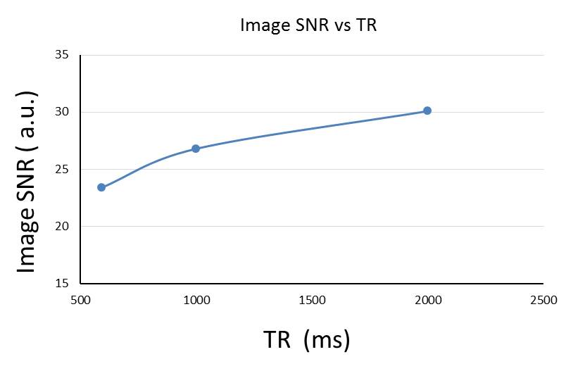
Figure.1
Changes of the signal to noise ratio (SNR) of the EPI images of monkey brains
vs different TR.
Images were acquired using multiband EPI sequences with TR = 590ms, 1000ms,
2000ms, and TE = 25 ms, MB
factor = 4.
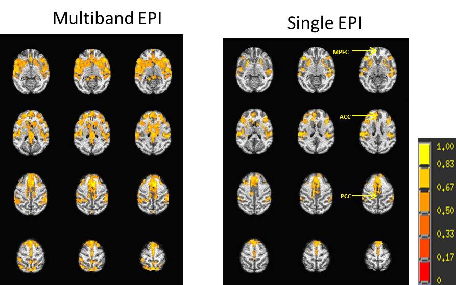
Figure.2
The correlation
maps (z maps) of default
mode network from multiband EPI (left, TR=1059ms,
TE=25ms, MB factor = 2)
and regular
single shot EPI
(right, TR=2190ms,
TE=25ms) at
same acquisition time (10 minutes) from a monkey. The
color bar represents the magnitude of the regression coefficient (z-score
threshold p<1×10-40 plus
cluster threshold 169 mm3
/overall. MPFC, medial prefrontal cortex; ACC,
anterior cingulate cortex; PCC, posterior cingulate cortex.
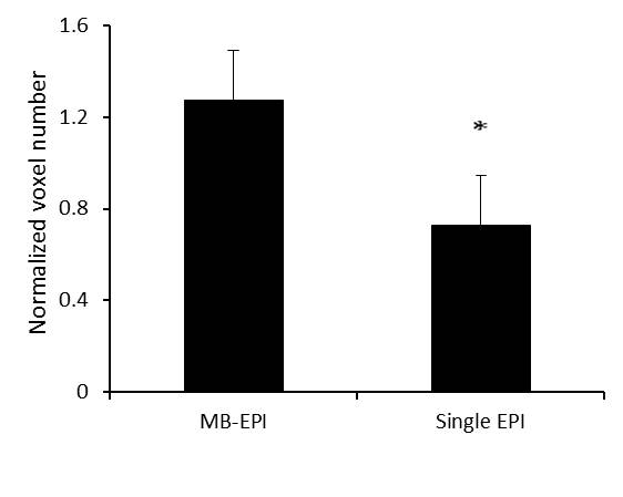
Figure.3
the normalized total voxel numbers of the normalized correlation maps across
default mode network (DMN) using multiband EPI (TR = 1059ms)
and regular EPI acquisition (TR = 2190ms) in anesthetized macaque monkeys.
The bar represents normalized
voxel numbers (normalized by average voxel numbers across DMN from two EPI acquis ion, z-score
threshold p<8×10-18 plus
cluster threshold 169 mm3
/overall, mean±SD.
n=4.*p=0.08,
paired t test. MB, multiband.