Kyung Eun Jang1, Yang-Tae Kim2, Jingu Kim3, Hyunsil Cha1, Heajung Choi1, Eunji Kim1, Moojin Yang1, Jiung Yang1, Huijin Song4, Moon Jung Hwang5, and Yongmin Chang1,6
1Medical & Biological Engineering, Kyungpook National University, Daegu, Republic of Korea, 2Psychiatry, School of Medicine, Keimyung University, Daegu, Republic of Korea, 3Physical Education, Kyungpook National University, Daegu, Republic of Korea, 4Biomedical Engineering Research, Kyungpook National University, Daegu, Republic of Korea, 5GE Health Korea, Seoul, Republic of Korea, 6Radiology and Molecular Medicine, College of Medicine, Kyungpook National University, Daegu, Republic of Korea
Synopsis
We
investigate neural activation of physical exercise related pictures in exercise
addiction. Our results demonstrate that exercise addiction group showed lower
activation in ventral striatum than the moderate exercise group, indicating
that dopamine release of the ventral striatum in exercise addiction group may
reduce because of withdrawal symptoms and negative prediction error. Moreover,
we found that exercise addiction group showed
less activation in the posterior
orbitofrontal cortex than the other groups, suggesting that exercise
addiction group may not deliberate fitness equipment and body shape of
exerciser as reward value.
Introduction
Exercise addiction is part of behavioral addictions with symptoms including craving, loss of control, withdrawal, and tolerance. It has been suggested that common neurobiological mechanism underlying substance and behavioral addiction may involve the reward circuit, including the ventral striatum and the orbitofrontal cortex [1]. The neural mechanism of exercise addiction in the perspective of reward circuit is still poorly understood. Therefore, the aim of this study was to investigate the change of activation in reward circuit of the ventral striatum and the posterior orbitofrontal cortex in exercise addiction.
Methods
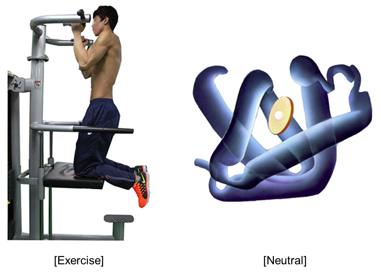
Seventy-seven right-handed healthy controls (22.8±2.3years, M:64, F:19) were included in this study. Participants were classified into three groups including exercise addiction, over-exercising, and moderate exercise group according to the Korean Exercise Addiction Scale. Each participant was presented physical exercise or neutral pictures during the fMRI acquisition (Figure 1). All fMRI data were acquired with a 3.0T MRI scanner (Discovery 750w; GE Healthcare). BOLD functional images were obtained using a single shot T2*-weighted gradignt EPI pulse sequence (TR=2000ms, TE=30ms, FOV=23cm, Matrix=64x64, Slice thickness=4.0mm, no gap, 32 contiguous axial slices, total scan time=6m8s). Image processing and statistical analysis for fMRI data were performed using SPM8 on MATLAB. The SPM{t} was thresholded at P<0.05, FWE-corrected for multiple comparisons across the whole brain for Monte Carlo simulation. Pearson correlation analysis were performed using SPSS software. Statistical significance was defined at P < 0.05.
Results
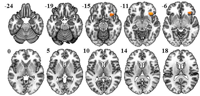
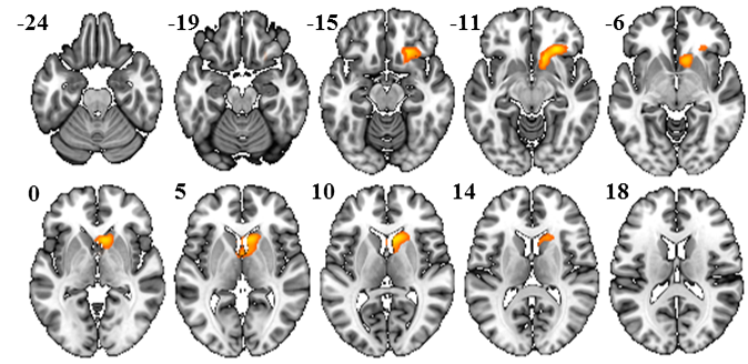
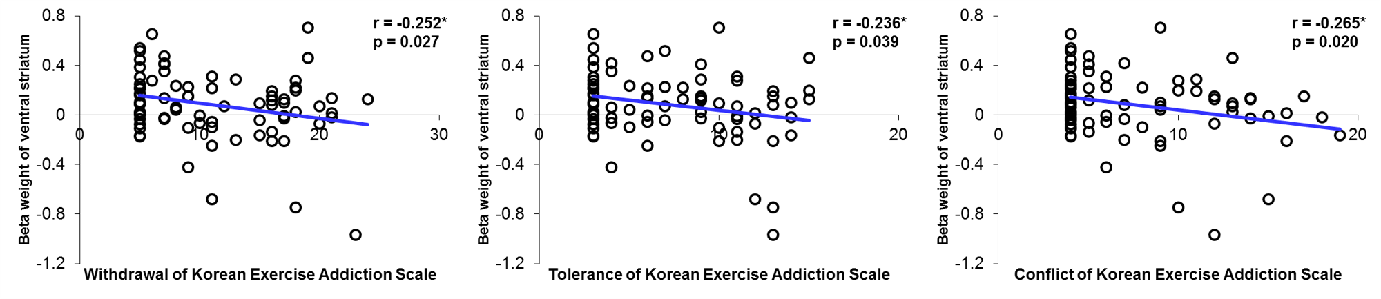
Exercise addiction group showed lower activation in ventral striatum than moderate exercise group (Figure 2). Furthermore, exercise addiction group showed less activation in posterior orbitofrontal cortex than the others group (Figure 3). In particular, exercise addicts showed higher scores than other groups in withdrawal, tolerance, and conflict of the Korean Exercise Addiction Scale. In addition, the activity of ventral striatum in exercise addicts has negative correlation with scores of withdrawal, tolerance, and conflict (Figure 4).
Discussion
We found that exercise addiction group showed less activation in ventral striatum and posterior orbitofrontal cortex than over-exercising group or moderate exercise group. The activation of ventral striatum in exercise addicts showed less than moderate exercise group. Specifically, the activity of ventral striatum has negative correlation with scores of withdrawal, tolerance, and conflict with Korean Exercise Addiction Scale. These findings may suggest two possible interpretation such as withdrawal symptoms and reward prediction. First, less activation of ventral striatum in exercise addiction group is related to withdrawal symptoms of addiction. Withdrawal of substance abuse in humans includes with fatigue, decreased motivation, and psychomotor retardation, which may be associated with the decrease in dopaminergic function [2]. Second, when exercise addicts watch a lower exerciser in pictures of physical exercise, the exercise addiction group felt less reward prediction. We also found that both over-exercising and moderate exercise group showed increased activation in the posterior orbitofrontal cortex than exercise addiction group. This finding suggests that exercise group with no addiction may consider various stimuli including body shape of exerciser and fitness equipment as reward value.
Conclusion
In the
present study, we investigated the neural mechanism of exercise addiction,
using fMRI. Our results may suggest that function of reward circuit in exercise
addiction could be hypoactive. This
study provides first evidence to elucidate neural mechanism in exercise
addiction.
Acknowledgements
No acknowledgement found.References
[1] Grant JE,
et al. Introduction to behavioral addictions. Am J Drug Alcohol Abuse (2010)
36(5):233-41.
[2]
George O, et al. Allostasis and addiction: role of the dopamine and
corticotropin-releasing factor systems. Physiol Behav. (2012) 106(1):58-64.