2372
Brain activity during the training period of the Hybrid Assistive Limb (HAL) for a subacute stroke: an fMRI case report1Center for Cybernics Research, University of Tsukuba, Tsukuba, Japan, 2Department of Neurosurgery, Ibaraki Prefectural University of Health Sciences Hospital, Ami, Japan, 3Department of Neurosurgery, Faculty of Medicine, University of Tsukuba, Tsukuba, Japan, 4Faculty of Engineering, Information and Systems, University of Tsukuba, Tsukuba, Japan, 5Department of Orthopaedic Surgery, Faculty of Medicine, University of Tsukuba, Tsukuba, Japan
Synopsis
The effectiveness of hybrid assistive limb (HAL) training, which is the new rehabilitation robotic approach, for recovery of brain function after stroke remains to be clarified. This is the first report to show the brain activation alteration during the training period for HAL for subacute stroke by using motor task-based functional magnetic resonance imaging (fMRI) 4 times. Our major finding was that fMRI results demonstrated rearrangement of the cortical activation pattern in a form that induces cerebral lateralization in M1 toward the contralateral hemisphere.
INTRODUCTION
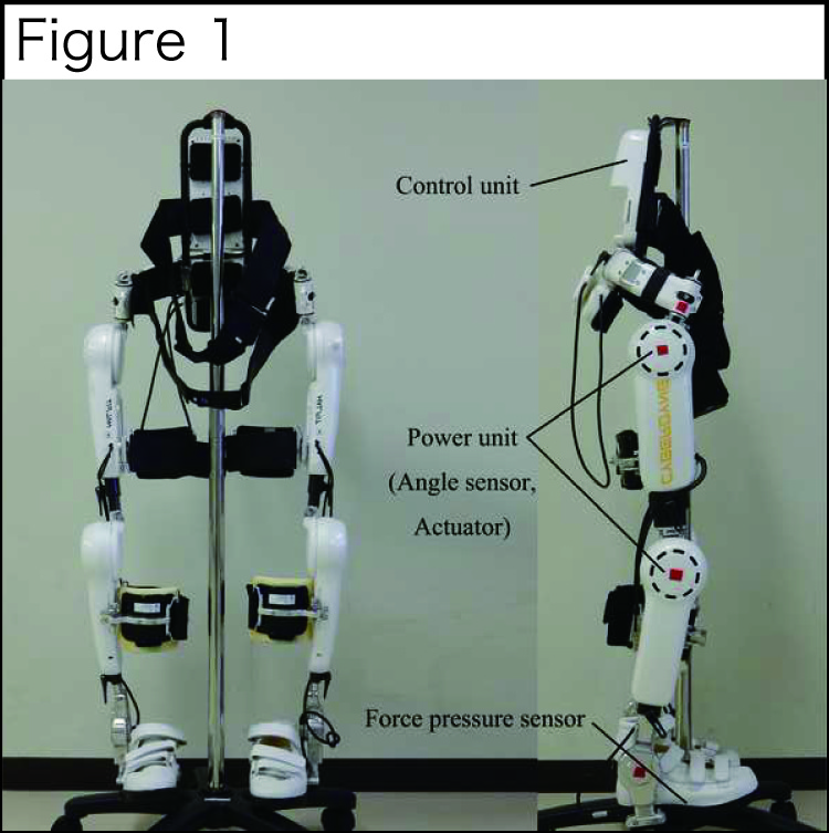
Hybrid Assistive Limb (HAL; Cyberdyne Inc, Japan) is a new rehabilitation robotic approach (Fig 1).1 The feasibility of HAL training for stroke patients should be evaluated. 2,3 This is the first report on changes in brain activation patterns in the primary motor cortex (M1) during training for HAL for subacute stroke by using motor task-based functional magnetic resonance imaging (fMRI) 4 times.CASE PRESENTATION
A 55-year-old woman had an atherothrombotic brain infarction in the right anterior cerebral artery 17 days before study entry. Neurological examination showed paralysis of the left ankle and foot. She received HAL training 3 times per week for 3 weeks (9 sessions, 40 minutes per session) with standard physical therapies. 10-m walk velocity and 6-minutes walk distance, and Fugl-Mayer Assessment of lower-extremity (FMA-L) score were measured after each HAL trainings.fMRI ACQUISITION
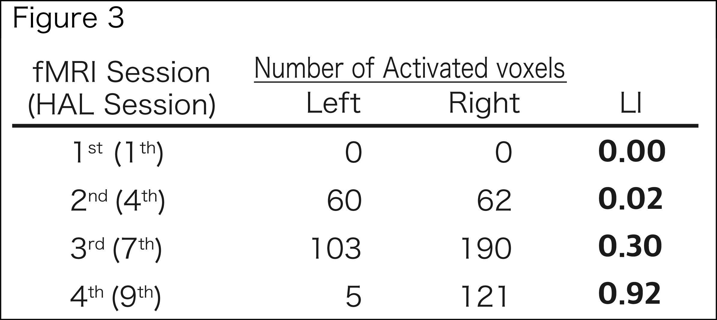
fMRI was performed 4 times during training for HAL (sessions 1, 4, 7, and 9), using a 3-T Philips Ingenia MRI system with echo planar imaging (2.5×2.5 mm in-plane resolution, TR/TE=2500/35 ms, 40 axial slices). Three-dimensional T1-weighted sequence was obtained (TR/TE=7.6/3.6 ms, 0.8×0.8 mm in-plane resolution). The patient performed a motor task of the left ankle movement with a block paradigm. Preprocessing included slice-time correction, realignment, de-spiking, and spatial normalization to MNI space using SPM12. A first-level fMRI model was constructed for the motor sequences, and contrast modeled the linear increase in BOLD signal due to increasing sequence difficulty levels. Region-of-interest analysis was applied to focus on the motor function of the precentral gyrus (PrG). We used the mask Right-Left PrG with SPM12 and a normalized index (laterality index [LI]) to quantify any shift in the cortical activation balance in the PrG during the training period of HAL.4 We calculated LI for the PrG in 4 sessions. LI is defined as LI=(R−L)/(R+L), where R and L are the active voxel count for the specified region in the right and left hemispheres, respectively. Then, we evaluated the correlation between LI and FMA-L score by using the Spearman correlation coefficient (R2).RESULTS
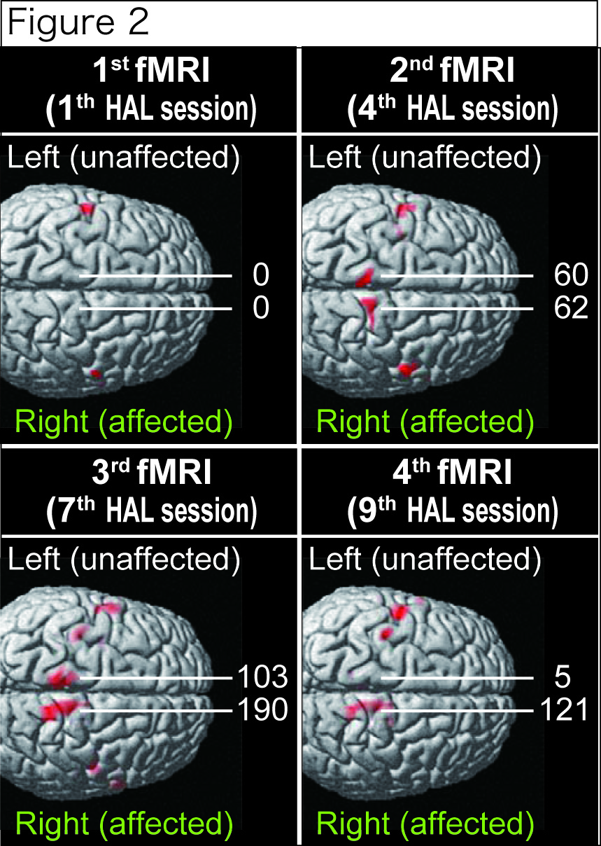
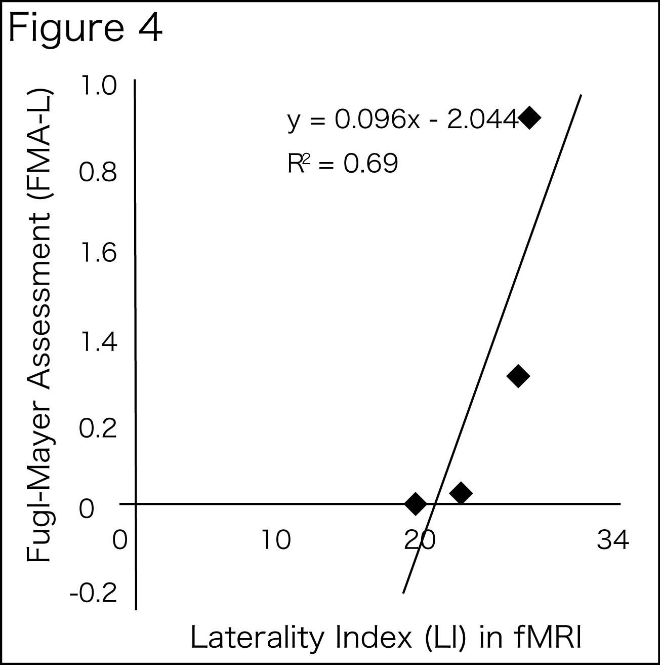
10-m walk velocity and 6-minutes walk distance increased from 31.3 m/s and 81.7 m to 41.2 m/s and 317.3 m. The FMA-L score was 20 points before HAL training and 27 points after HAL training. Figure 2 shows that the activation maps for the right and left PrG masked the ROIs at each HAL session. Decreased signal was present in the left side (ipsilateral) but not in the right side (contralateral). The LIs for the PrG regions at the HAL sessions were 0.00, 0.02, 0.30, and 0.92, respectively (Figure 3). LI significantly correlated with FMA-L score (R2=0.69, P<0.01; Fig 4).DISCUSSION
Some studies focused on the changes in measures related to motor ability to clarify the clinical potential of HAL for chronic or acute stroke.4 However, the effectiveness of HAL training for recovery of brain function after stroke remains to be clarified. Our major finding is that fMRI results demonstrated rearrangement of the cortical activation pattern, inducing cerebral lateralization in M1 toward the contralateral hemisphere. Although generalization from the results of a single-case study should be made with caution, this suggests that HAL training could drive neuroplasticity in the cerebral cortex in patients with subacute stroke. LI positively correlated with FMA-L score, which suggests that lean locomotor activity due to repetitive generation of voluntary movement using HAL improved the ability of unilateral ankle movement and subserved the perception of spatial position of the left ankle, allowing a shift to the contralateral hemisphere in activation balance. Considering that all fMRI acquisitions were performed after training, these fMRI results might show a longitudinal change in the immediate effect due to HAL. More cases and further controlled studies are required to clarify the changes in brain function associated with HAL training for strokes.CONCLUSION
4 times motor task-based fMRIs during the training period for HAL for patients with an atherothrombotic brain infarction in the right anterior cerebral artery 17 days before study entry were performed. Long-lasting voluntary locomotor activity using a HAL device could drive neuroplasticity in the cerebral cortex for patients with subacute stroke.Acknowledgements
This study was supported by the Industrial Disease Clinical Research Grants of the Ministry of Health Labour and Welfare, Japan (14060101-01) and funded by ImPACT Program of Council for Science, Technology and Innovation (Cabinet Office, Government of Japan).References
1. Kawamoto H, Sankai Y. Power assist method based on Phase Sequence and muscle force condition for HAL. Adv Robot. 2005;19:717-34.
2. Wall A, Borg J, Palmcrantz S. Clinical application of the Hybrid Assistive Limb (HAL) for gait training-a sustematic review. Front Syst Neurosci. 2014;9:48.
3. Ueba T, Hamada O, Ogata T, Inoue T, Shiota E, Sankai Y: Feasibility and safety of acute phase rehabilitation after stroke using the hybrid assistive limb robot suit. Neurol Med Chir 2013, 53:287–290.
4. Cramer SC, Nelles G, Benson RR, et al. A Functional MRI Study of Subjects Recovered From Hemiparetic Stroke. Stroke. 1997;28:2518-2527.
Figures