2362
Visual cortex and auditory cortex activation in early binocularly blind macaques: A BOLD-fMRI study using auditory stimuliLingjie Wu1, Rong Wang2, Zuohua Tang2, Xinghuai Sun3, Xiaoyuan Feng4, Weijun Tang4, Wen Qian2, Jie Wang1, Yufeng Zhong5, Zebin Xiao2, and Zhongshuai Zhang6
1Otolaryngology, Eye & ENT Hospital of Fudan University, Shanghai, China, 2Radiology, Eye & ENT Hospital of Fudan University, Shanghai, China, 3Eye, Eye & ENT Hospital of Fudan University, Shanghai, China, 4Radiology, Huashan Hospital of Fudan University, Shanghai, China, 5Jinshan Hospital of Fudan University, Shanghai, China, 6Siemens Healthcare Ltd., Shanghai, China
Synopsis
We aimed to detect the changes in BOLD activity between the visual and auditory cortices of monocularly blind neonatal macaques by using pure tones as auditory stimuli. The changes in the BOLD response in the bilateral visual and auditory cortices were detected and further compared with the findings of the immunofluorescent staining. In monocularly bind macaques, we found a greater level of significant activation in the bilateral visual cortices while the number of activated volumes of the bilateral auditory cortices decreased. Therefore, cross-modal plasticity within the visual and auditory cortices was established in the monocularly blind macaques.
Purpose
Although blind individuals lose visual function, their non-visual functions (such as the senses of touch, hearing and smell) are more enhanced than those of sighted people [1-2]. This phenomenon may be because the visual cortex can be involved in processing non-visual information through a new channel between the visual cortex and other sensory cortices, which is called cross-modal plasticity [3-4]. blood-oxygen-level-dependent functional magnetic resonance imaging (BOLD-fMRI) has become an effective tool for detecting the functional reorganization of the visual cortex after vision loss [5]. In this study, we aimed to investigate cross-modal plasticity within the visual and auditory cortices of macaques with early monocular blindness by using BOLD-fMRI combined with immunofluorescence staining.Materials and methods
Four healthy neonatal macaques (postnatal day 7) were randomly divided into 2 groups (group A and group B, 2 macaques in each group). The macaques in group A were kept as a control group. Optic nerve transection was performed in the right eye of the macaques in group B to establish the monocularly blind model. Sixteen months later, all macaques underwent a BOLD-fMRI scanning protocol involving pure tone auditory stimuli. Data were collected on a 3T MR scanner (MAGNETOM Verio, Siemens Healthcare, Erlangen, Germany) with a 32-channel knee joint coil. T1-weighted images were acquired for each macaque using a magnetization-prepared rapid gradient echo (MPRAGE) sequence to obtain anatomical information (TR=500 ms, TE=12 ms, matrix=128×128, FOV=102×102 mm2, slice thickness=2 mm, averages=4). BOLD-fMRI data were acquired through an echo planar imaging (EPI) sequence (TR=3000 ms, TE=30 ms, matrix=128×128, FOV=192×192 mm2, 80 axial slices, slice thickness=2 mm, gap between slices=1 mm, averages=1).Results
Compared with group A, significant activation is observed via BOLD-fMRI in the bilateral visual cortices of the monocularly blind macaques in response to the auditory stimuli. To be more specific, they are more obvious in the left visual cortices compared to those in the right (Figure 1). These findings are consistent with the results of the immunofluorescence staining, which showed that the number of c-Fos-positive cells in the bilateral visual cortices of monocularly blind macaques is greater than that in the normal macaques (p<0.01) and this phenomenon is more obvious in the left visual cortices (Figure 2). In addition, we also find less activated volumes in the bilateral auditory cortices in monocularly blind macaques than those in normal macaques. These findings are also in line with the results of the immunofluorescence staining, which show a smaller number of c-Fos-positive cells in the bilateral auditory cortices of the monocularly blind macaques than in those of the normal animals (p<0.05), particularly in the left auditory cortices (Figure 2). Furthermore, we find that the frontal cortex is also activated in monocularly blind macaques in response to the auditory stimuli of pure tones (Figure 1).Discussion
As a result of decreased activation in the auditory cortex, the visual cortex of monocularly blind macaques could be activated in response to auditory stimuli; these findings are confirmed by c-Fos immunofluorescence staining, which indicates that cross-modal plasticity can be established within visual and auditory cortices. In addition, the frontal cortex can also be activated in order to process auditory stimuli to further compensate for the vision loss in these macaques.Acknowledgements
Sources of support that require acknowledgment: The Commission of Shanghai Municipality (No.09ZR1405600, No.09JC1403100, No.14411962000); The Funds for International Cooperation and Exchange of the National Natural Science Foundation of China (Grant No. 81020108017).References
[1]Beaulieu-Lefebvre, M., Schneider, F.C., Kupers, R., Ptito, M. (2011) Odor perception and odor awareness in congenital blindness. Brain Research Bulletin 84, 206-209. [2]Wolbers, T., Zahorik, P., Giudice, N.A. (2011) Decoding the direction of auditory motion in blind humans. Neuroimage 56, 681-687. [3]Anurova, I., Renier, L.A., Volder, A.G.D., Carlson, S., Rauschecker, J.P. (2015) Relationship Between Cortical Thickness and Functional Activation in the Early Blind. Cerebral Cortex 25, 2035. [4]Qin, W., Xuan, Y., Liu, Y., Jiang, T., Yu, C. (2015) Functional Connectivity Density in Congenitally and Late Blind Subjects. Cerebral Cortex 25, 2507. [5]Sabbah, N., Sanda, N., Authié, C.N., Mohandsaïd, S., Sahel, J., Habas, C., Amedi, A., Safran, A.B. (2017) Reorganization of early visual cortex functional connectivity following selective peripheral and central visual loss. Scientific Reports 7, 43223.Figures
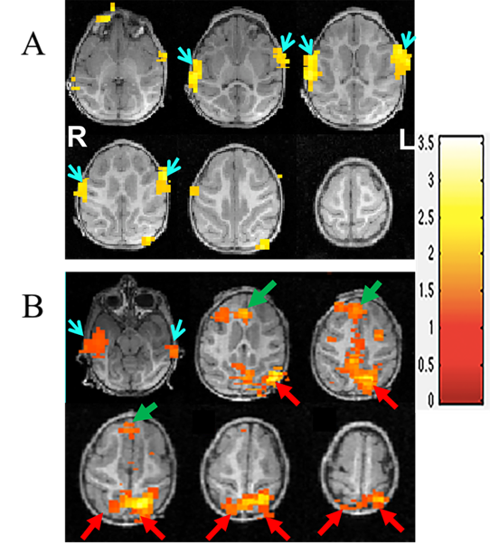
The brain activation map of the normal (A) and monocularly blind macaques
(B) in response to the auditory stimuli produced using BOLD-fMRI. The
bilateral visual cortices of the monocularly blind macaques are significantly
activated (red arrows), especially in the left visual cortex (B), whereas the
bilateral visual cortices of the normal macaques are not activated (A). Moreover,
less activated volumes are observed in the auditory cortices (blue arrowheads) of
the monocularly blind macaques than in those of the normal macaques (blue arrowheads).
In addition, the frontal cortex
(green arrows) is also activated in these macaques.

C-Fos
immunofluorescence staining of the bilateral visual cortices (A1, A2, B1, B2)
and bilateral auditory cortices (A3, A4, B3, B4) in normal and monocularly
blind macaques. The densities of the c-Fos-positive cells in the
left (B1) and right (B2) visual cortices of the monocularly blind macaques were
greater than those in the
normal macaques (A1, A2). In addition, the
densities of the c-Fos-positive cells in the bilateral auditory cortices (B3,
B4) were lower in the monocularly blind macaques than in the normal macaques
(A3, A4).
Inter-group and intra-group comparisons are shown as a histogram (C). Group A, control group; Group B,
monocularly blind group.