2358
BOLD responses in the posterior cerebellum differ when a motor task has a proprioception component1Spinoza Centre for Neuroimaging, Amsterdam, Netherlands
Synopsis
The cerebellum receives proprioceptive information from the body, as well as tactile input. Here, we aimed to separate the proprioceptive BOLD responses from the motor/somatosensory clusters in the human cerebellum. Regions responding to a fingertapping task and a motion task requiring proprioceptive information were found to differ in the posterior cerebellum. Using high resolution 7T functional MRI, all proprioceptive clusters in lobule VIII of the cerebellum were found to be positioned medial to regions responding to the simple tapping task.
Introduction
The cerebellum receives proprioceptive information from the body, as well as tactile input1. BOLD responses in the cerebellum are generally difficult to disentangle, primarily because the cerebellum is small and its cortex is thin and very highly folded. High spatial resolution is essential in this brain region. However, even with state-of-the art imaging and analysis techniques the cerebellar motor and somatosensory BOLD responses could not be separated, suggesting a degree of shared neural circuitry or at least intermingled responses in the cerebellum2. Here, we tested whether 7T fMRI could be used to distinguish related information, i.e. proprioceptive BOLD responses, from those associated with a tactile/motor-only task.
The purpose of this study was to separate cerebellar BOLD responses to tasks with and without a proprioceptive component using 7T fMRI.
Methods
Five volunteers were scanned at 7T (Philips, NL). Each performed two functional runs (3D-EPI, 1.3mm resolution, TRvol/TR/TE/α = 3s/44ms/26ms/16o, matrix size 156x156x31, SENSE factor 3 (RL), 155 volumes) with a motor task alternating blocks of 15s task with 15s rest. During task blocks participants were either instructed to move the same digits on each hand (e.g. move both thumbs) in a simple motor task or to touch two fingers over their abdomen (e.g. left thumb touches right middle finger) in a more challenging “proprioceptive” task. Instructions changed every 2 seconds. Proprioceptive information is required to execute this task because participants were not able to see their hands while laying supine in the scanner bore. Because the digits were touching, both tasks involved equal amounts of tactile stimulation as well as a comparable motor component. Careful positioning of the lower arms was necessary to avoid arm movements in the two-handed proprioceptive task. T1-weighted 3D-EPI data3 were also acquired to aid coregistration to the anatomical MP2RAGE4. Dielectric pads were used to improve B1 homogeneity over the cerebellum5. Five EPI volumes were acquired with a reversed phase-encoding direction to allow distortion correction using FSL’s TOPUP routine.
Data analysis: All data were analysed in SPM using a GLM analysis following motion correction and minimal spatial smoothing (FWHM 2mm). Besides the movement regressors temporal derivatives were included to compensate for lags in the onset of movement following the display of the stimulus. An individual subject analysis approach was chosen to retain spatial resolution.
Results
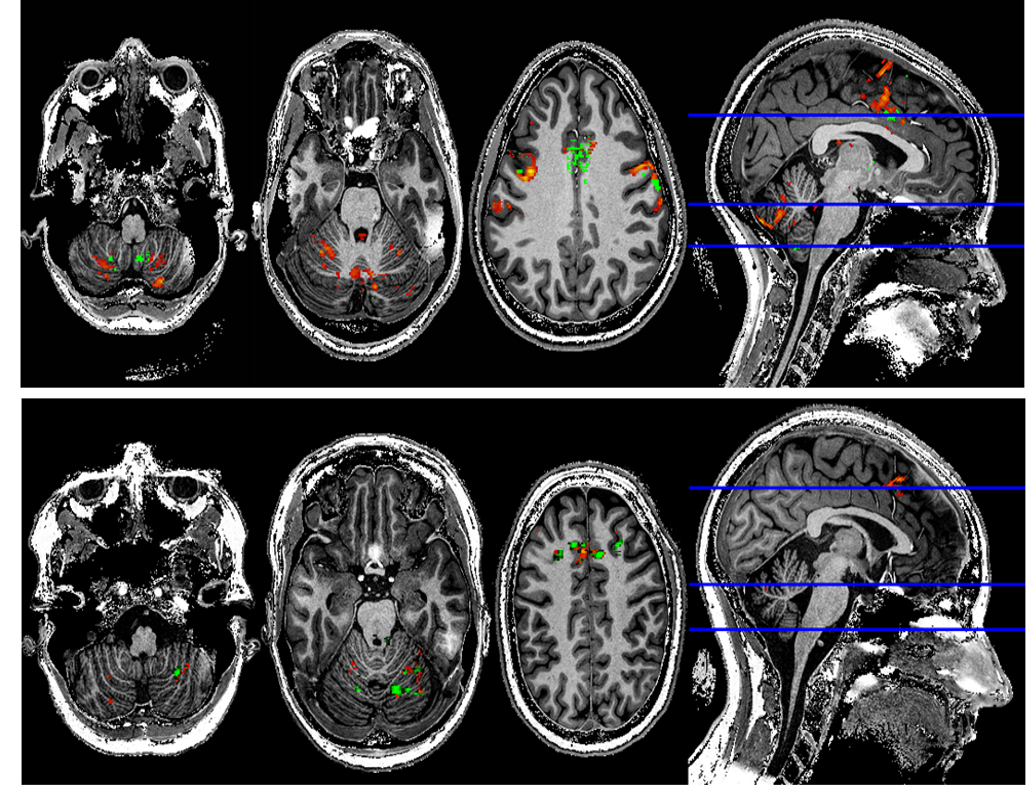
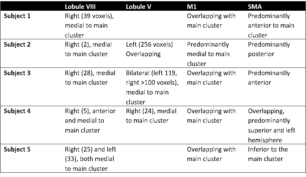
BOLD responses (p<0.05 FWE) associated with the pointing task were detected in all subjects bilateral in the cerebellar lobules V (CV) and VIII (CVIII), as well as in the supplementary motor area (SMA) and bilaterally in the primary motor cortex (MI). In the cerebellum, small regions just medial or anterio-medial to these clusters showed significantly larger responses to the “proprioceptive” task (p<0.001, uncorrected) in lobule V (3/5 subjects), but especially in lobule VIII (5/5) (See Figure 1 and Table 1). These proprioceptive clusters were found predominantly, but not exclusively, in the right cerebellar hemispheres. Subregions of the cerebral motor regions also showed larger responses to the proprioceptive task, but in these no consistent spatial pattern was found (Table 1).Discussion
The individual subject analysis, spatial resolution and high BOLD sensitivity at 7T were essential to find the small clusters surpassing the significance threshold for the proprioceptive > tapping contrast. Clusters were usually separated from the tapping>rest clusters by only some 5-10 millimetres. The minimal spatial smoothing did not impact the detection of these clusters, but might obscure BOLD responses located even closer together. An alternative to spatial smoothing to improve detection power would be group averaging. The high inter-subject variability in gross cerebellar anatomy impedes straightforward normalisation in a 3D space, which could however be undertaken in a cerebellar cortical space, given anatomical data of sufficient quality to fully segment the cortex.
Residual arm movements in the proprioceptive task might also lead to differing BOLD responses compared to the motor-only task, but hand/arm movements are known to elicit BOLD responses in the same parasagittal plane as the digit oppositions used in the motor-only run6. BOLD responses for hand/arm movements are hence not expected in the more medial plane in which the proprioceptive responses in this experiment were found.
Conclusion
Regions responding to a fingertapping task and a motion task requiring proprioceptive information were found to differ in the posterior cerebellum. Using 7T fMRI, all proprioceptive clusters in lobule VIII of the cerebellum were found to be positioned medial to regions responding to the simple tapping task.Acknowledgements
The author would like to thank Alessio Fracasso and Pierre-Louis Bazin for helpful discussions on cerebellar fMRI in general and data analysis in particular.References
1. Manto, M. et al. Consensus Paper: Roles of the Cerebellum in Motor Control—The Diversity of Ideas on Cerebellar Involvement in Movement. Cerebellum Lond. Engl. 11, 457–487 (2012).
2. Wiestler, T., McGonigle, D. J. & Diedrichsen, J. Integration of sensory and motor representations of single fingers in the human cerebellum. J. Neurophysiol. 105, 3042–3053 (2011).
3. van der Zwaag, W., Buur, P. F., Versluis, M. & Marques, J. P. Distortion-matched T1maps and bias-corrected T1w images as anatomical reference for submillimeter-resolution fMRI. ISMRM 2016,
4. Marques, J. P. et al. MP2RAGE, a self bias-field corrected sequence for improved segmentation and T1-mapping at high field. NeuroImage 49, 1271–1281 (2010).
5. O’Brien, K. R. et al. Dielectric pads and low- B1+ adiabatic pulses: complementary techniques to optimize structural T1 w whole-brain MP2RAGE scans at 7 tesla. J. Magn. Reson. Imaging JMRI 40, 804–812 (2014).
6. Van der Zwaag, W. et al. Digit somatotopy in the human cerebellum: A 7T fMRI study. NeuroImage 67, 354–362 (2013).
Figures