2354
Changes in functional connectivity in the ventral attention system1Department of Radiology, Centre Hospitalier Universitaire Vaudois (CHUV), and University of Lausanne, 1011 Lausanne, Switzerland, Unil, Lausanne, Switzerland, 2Neuropsychology and Neurorehabilitation Service, Centre Hospitalier Universitaire Vaudois (CHUV), and University of Lausanne, 1011 Lausanne, Switzerland, Unil, Lausanne, Switzerland, 3CIBM (Centre d'Imagerie Biomédicale), Dept. of Radiology, Centre Hospitalier Universitaire Vaudois (CHUV), and University of Lausanne, 1011 Lausanne, Switzerland, Unil, Lausanne, Switzerland
Synopsis
Prismatic adaptation (PA), a therapy for neglect after a stroke, showed conflicting results in the literature. The variability of brain lesions leading to neglect and their relation to structures affected by PA could explain those results. MRI studies found focal brain activations after PA. We aim to understand if there is an effect on functional networks after PA. To do so, we analyzed the resting state fMRI connectivity of healthy subjects before and after PA. We found that the ventral attention system (VAS) was less connected after PA, this provides new insights to select patients for this therapy.
Introduction and purpose
Prismatic adaptation (PA) consists of a brief session during which subjects point to targets under visual control while wearing goggles with prisms that deviate the visual field1. Such adaptation is used in the rehabilitation of spatial neglect, a common disorder following right hemispheric damage, characterized by disturbances of spatial representation and attention, with a tendency to ignore stimuli in the left visual field2. In control subjects, task-related fMRI have shown that PA modulates principally the activity of the inferior parietal lobule (IPL)3,4 suggesting that PA changes the right hemispheric dominance within the Ventral Attention System (VAS) by shifting the visual field representation from right to left IPL hemisphere3. Here, we tested this hypothesis by using resting-state fMRI acquisition before and after a PA session and investigated PA-related modulations of the VAS, the Dorsal Attention System (DAS) and the Default Mode Network (DMN).Method
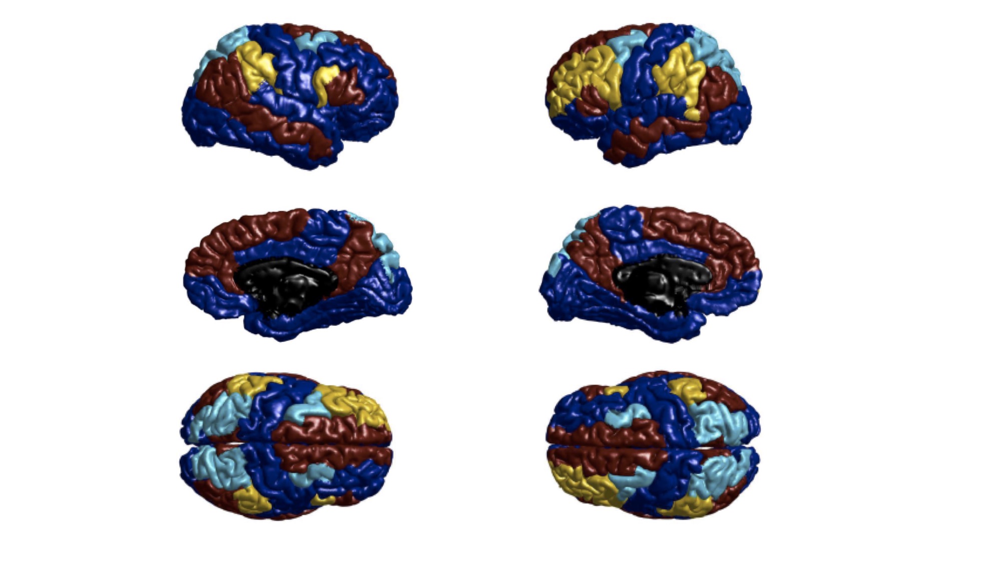
We obtained on 26 subjects 10 minutes of resting state functional MRI data before and after an adaptation (3T Siemens Magnetom Trio scanner with a 32-channel head-coil, TR=2sec, 32 slices, 3mm isotropic). During the adaptation 14 subjects wore rightward deviating prisms (treated group), 12 other subjects wore plain glasses (control group). Graph metrics on resting state connectivity matrices were calculated using Matlab and the brain connectivity toolbox5. We worked with a brain parcellation of 83 regions of interest (ROIs)6 and focused on three networks, the VAS, DAS and DMN (Fig. 1). After a consultation with an expert, we selected the ROIs of the aforementioned networks based on the Yeo 7 functional systems parcellation7. For each network separately, the mean correlation coefficient of connections between nodes of the network, called total connectivity5, was calculated. The differences ‘before-after adaptation’ were compared between groups using bilateral independent-samples t-tests in SPSS.Results
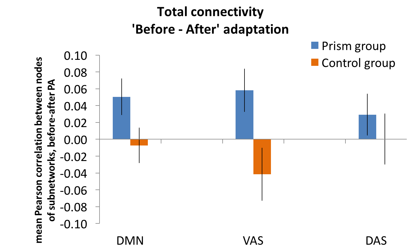
The total connectivity was lower after compared to before adaptation for the DMN, VAS in the prisms group and for the DAS in both groups, whereas it was higher after compared to before adaptation only for the control group in the DMN and VAS (Fig. 2). However, the difference between groups was significant only for the VAS network (t(24)=2.49;p=0.02), not for the DMN (t(24)=1.90;p=0.69) nor the DAS (t(24)=0.75;p=0.461).Discussion and conclusion
Our results provide new evidence for a modulation of the attentional systems by rightward PA. More precisely, PA changes significantly the functional connectivity in the VAS, and neither in the DAS nor in the DMN. The VAS, including the temporo-parietal junction and the ventral frontal cortex, responds typically to behaviorally relevant stimuli occurring unexpectedly, as opposed to the DAS that includes the intraparietal sulcus and frontal eye fields and underlies active orientation of attention8. Our findings confirm that, as proposed in the SHD-VAS model9, PA modulates the ventral attentional regions. Taken together with previous findings showing an increased activation in the left IPL after PA, our current results suggest that PA uncover spatial representation of ventral regions of the left hemisphere by changing the connectivity in the VAS. Our results provide new insights to optimize the choice of PA for the rehabilitation of spatial neglect for patients having a spared VAS.Acknowledgements
No acknowledgement found.References
1. Rossetti, Y. et al. [Prism adaptation to a rightward optical deviation rehabilitates left hemispatial neglect]. Nature 395, 166–169 (1998).
2. Parton, A., Malhotra, P. & Husain, M. Hemispatial neglect. J. Neurol. Neurosurg. Psychiatry 75, 13–21 (2004).
3. Crottaz-Herbette, S., Fornari, E. & Clarke, S. Prismatic Adaptation Changes Visuospatial Representation in the Inferior Parietal Lobule. J. Neurosci. 34, 11803–11811 (2014).
4. Crottaz-Herbette, S. et al. Reshaping the brain after stroke: The effect of prismatic adaptation in patients with right brain damage. Neuropsychologia 104, 54–63 (2017).
5. Rubinov, M. & Sporns, O. Complex network measures of brain connectivity: Uses and interpretations. NeuroImage 52, 1059–1069 (2010).
6. Hagmann, P. et al. Mapping the Structural Core of Human Cerebral Cortex. PLoS Biol. 6, (2008).
7. Yeo, B. T. T. et al. The organization of the human cerebral cortex estimated by intrinsic functional connectivity. J. Neurophysiol. 106, 1125–1165 (2011).
8. Vossel, S., Geng, J. J. & Fink, G. R. Dorsal and ventral attention systems: distinct neural circuits but collaborative roles. Neurosci. Rev. J. Bringing Neurobiol. Neurol. Psychiatry 20, 150–159 (2014).
9. Clarke, S. & Crottaz-Herbette, S. Modulation of visual attention by prismatic adaptation. Neuropsychologia 92, 31–41 (2016).
Figures