2342
Research on the brain function of cervicogenic vertigo: A resting-state fMRI study1Radiological Department, Renmin Hospital of Wuhan University, Wuhan, China, 2GE Healthcare China, Beijing, China
Synopsis
People with cervicogenic vertigo(CV) due to vertebrobasilar insufficiency suffer lots of troubles. Through neuroimaging analysis method, this study finds significant difference in right cerebellum anterior lobe(RCAL) on mfALFF value and connectivities with other brain regions between CV and normal control(NC). Besides, the mfALFF and mReHo of RCAL are correlated to DHI(Dizziness Handicap Inventory) significant positively. These discoveries seem to indicate that a long-term vertebrobasilar insufficiency results in such alterations of these functional indexes and connectivities in RCAL of CV, then lead to the function degradation of maintain the basic balance of the body when occurrence of vertigo.
Introduction
Recent studies1-3 indicate that the cervicogenic vertigo(CV) is due to vertebrobasilar insufficiency. However, those studies were only confined to investigate the structure and function of vertebral body and vertebral artery. The studies focused on formation and maintenance mechanism of vertigo in view of neurological are few. Through neuroimaging analysis method, this article investigates that whether a long-term vertebrobasilar insufficiency cause function alterations of some brain regions, then lead to the degradation of their corresponding function, to provide a new perspective for its clinical treatment.
Methods
According to the inclusion criteria of the research4, 29 patients with CV and 23 normal controls (NC) were recruited in this study. There was no significant difference in age and gender between the two groups. All MR images were acquired on a 3.0T MR scanner (GE Discovery MR 750), which include sagittal 3D T1 weighted Bravo images and resting-state BOLD fMRI images. The DHI (Dizziness Handicap Inventory) scale of CV group was tested. Pre-processing procedures and statistical analysis were conducted through SPM5, DPABI6, and CONN7. In two conditions of with and without global signal regression, the mfALFF and mReHo of two groups were calculated and tested with two sample T-test. The brain areas with accordant statistically alterations of both mALFF and mReHo values were treated as seeds for further analysis. The functional connectivities between seeds and the whole brain were analyzed and compared for two groups. Also, correlation analysis between the mfALFF and mReHo value of interest brain regions under the above two conditions with DHI score of CV group were conducted.Results
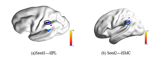
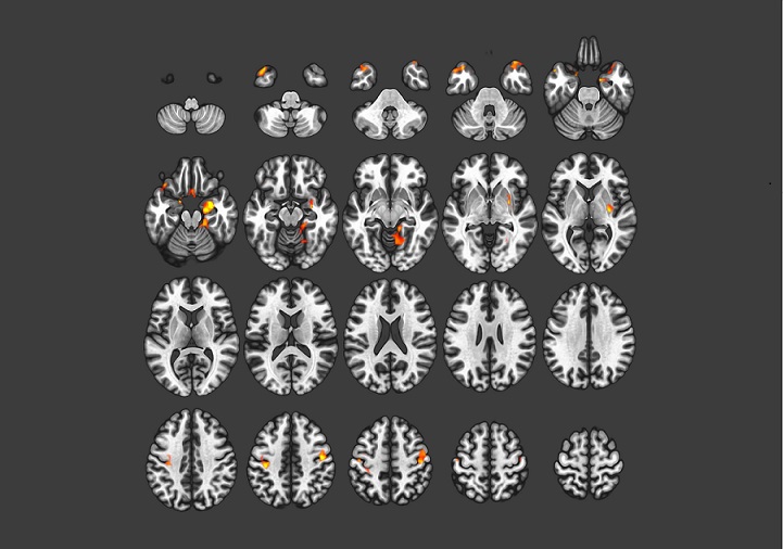
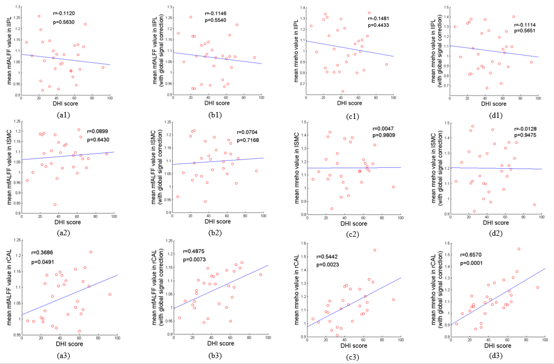
Whether the global signal was regressed or not, the mfALFF values in left inferior parietal lobule (lIPL) and left supramarginal cortex (lSMC) of CV are significantly decreased compared to the NC group (see Fig. 1 a and b). So were the mReHo values (see Fig. 1 c and d). Besides, the mfALFF values in right cerebellum anterior lobe (rCAL) of CV are significantly higher than NC. But there was no significant between group difference for mReHo value in this region. As a result, lIPL and lSMC were used as seed regions for further analysis (see Fig. 2). Taking lIPL as seed, the functional connectivity between seed and rCAL of CV is significantly increased compared to the NC group (see Fig. 3). However, there is no significantly between group functional connectivity difference when taking lSMC as seed. In correlation analysis, we take lIPL, lSMC and rCAL as interest brain regions. there were no significant correlation between mfALFF or mReHo in lIPL or lSMC with DHI score (see Fig.4(a1-d1), (a2-d2) ) . But, the DHI score is significantly positive correlated to mfALFF and mReHo in rCAL with global signal regressed or not (see Fig. 4(a3-d3) ).Discussion
lIPL and lSMC are affiliated to the default mode network. The mfALFF and mReHo in these two brain areas of CV are significantly lower than NC. To some extent, it indicates that the vertigo of CD accompanied by the function degradation of monitoring external environment executed by the default mode network. Besides, the mfALFF in rCAL of CV increase significantly. Whether it can be interpreted that a long-term vertebrobasilar insufficiency cause a few collateral circulation to initiate the abnormal activation of neurons in rCAL, so it is unable to perform its normal function to maintain the basic balance of the body. Thus, when the occurrence of vertigo, the body loses balance control ability. Furthermore, the connectivity between lIPL and rCAL of CV significantly increase. It seems that the abnormal mfALFF increase in rCAL of CV because of a few abnormal connections between it and other brain regions. Interestingly, significantly positive correlation between DHI with mfALFF and mReHo in rCAL with global signal regressed or not appear to show that with the progression of the disease the synchronous activity of neurons in rCAL unusually increased, caused its abnormal increase of mfALFF and connections with other brain regions. These results seem to be complemently to each other. This study provides compensatory insight into the neural mechanisms for CV.Keywords
Cervicogenic vertigo, rest-fMRI, mfALFF, mReHo, connectivityAcknowledgements
No acknowledgement found.References
1.Yongchao Li, Baogan Peng. Pathogenesis, Diagnosis, and Treatment of Cervical Vertigo. Pain Physician. 2015;18:E583-E595.
2.Noh Y, Kwon OK, Kim HJ, Kim JS. Rotational vertebral artery syndrome due to compression of nondominant vertebral artery terminating inposterior inferior cerebellar artery. J Neurol 2011; 258:1775-1780.
3.Choi KD, Choi JH, Kim JS, Kim HJ. Rotational vertebral artery occlusion: mechanisms and long-term outcome. Stroke 2013; 44:1817-1824.
4.Susan A. Reid, Robin Callister, Michael, Julia M. Utility of a brief assessment tool developed from the Dizziness Handicap Invebtory to screen for Cervicogenic dizziness: A case control study. Musculoskeletal Science and Practice 2017;30:42-48.
5. Ashburner,J. SPM: a history. Neuroimage, 2012; 62(2), 791-800.
6 Yan, C.G., Wang, X.D., Zuo, X.N., Zang, Y.F.. DPABI: Data Processing & Analysis for (Resting-State) Brain Imaging. Neuroinformatics. 2016;14, 339-351.
7. Susan Whitfield, Alfonso Nieto. Conn: A Functional Connectivity Toolbox for Correlated and Anticorelated Brain Networks.Brain Connectivity. 2012;2(3):125-141.
Figures
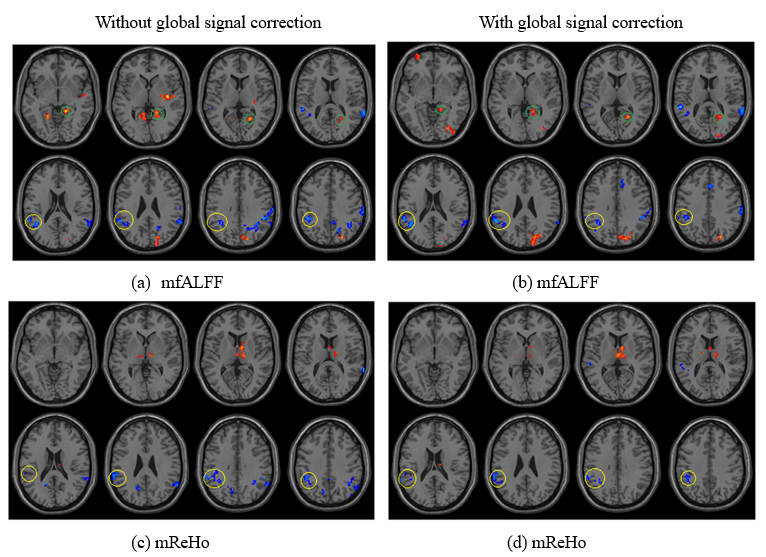
Figure 1. (a)(b)Two sample T-tests of mfALFF between two group in two conditions of without and with global signal regression(CV-NC,P<0.05,uncorrected);
(c)(d)Two sample T-tests of mReHo between two group in two conditions of without and with global signal regression (CV-NC,P<0.05,uncorrected).
Green circle--rCAL; Yellow circle--lIPL and rSMC.