2318
Multi-scale assessment of brain network response to sustained working memory task1Centro Fermi - Museo storico della fisica e Centro di studi e ricerche Enrico Fermi, Rome, Italy, 2Dipartimento di Neurologia e Psichiatria, Sapienza Università di Roma, Roma, Italy, 3Fondazione Santa Lucia IRCCS, Roma, Italy, 4Dipartimento di Fisica, Sapienza Università di Roma, Roma, Italy, 5Cardiff University Brain Research Imaging Centre (CUBRIC), Cardiff, United Kingdom, 6Center for Magnetic Resonance Research, University of Minnesota, Minneapolis, MN, United States, 7ImpAct Team, Lyon Neuroscience Research Center, Lyon, France
Synopsis
How low-frequency BOLD fluctuations (LFFs) are modulated when the brain is engaged in processing external stimuli is still poorly described. We exploited a non-conventional, long-lasting, block-design paradigm to study LFF modulations during sustained performance of a working memory task. Task-associated modulations were characterized by increased synchronization between networks at the expense of reduced within-network coherence. Such pattern persisted at several spatial scales, indicating a scale-invariant feature of task-associated modulations. Despite such clear-cut network behavior, no linear correlation between performance and connectivity changes was observed. Contrarily, high levels of connectivity at task and especially at rest were associated with greater performance.
INTRODUCTION
Low-frequency BOLD fluctuations (LFFs) have been extensively studied during resting-state conditions, leading to the robust identification of several brain networks characterized by synchronous oscillations1. While LFFs are known to persist during task execution2, how they are modulated during sustained stimulations, as well as the behavioral relevance of their modulation, is still matter of debate. Here we exploited a working memory (WM) block-design paradigm composed of long-lasting epochs (~5 minutes) to properly disentangle the task-associated LFFs from the localized task-evoked BOLD response. The LFF component was examined to investigate 1) the functional connectivity (FC) modulations associated with the sustained WM task, 2) the spatial scale at which modulations happen, and 3) the behavioral relevance of such modulations.METHODS
Twenty-healthy subjects (12M/8F, 33 ± 6 y.o.) were scanned on a 3T Siemens Allegra. Two functional scans were collected using a 2D gradient EPI (TE/TR=30/2100ms, FA=70°, 3×3×2.5mm3 voxel size), covering the whole brain. Each scan lasted 24 minutes and 38 seconds and consisted of alternated epochs (4min and 54s each) of open-eyes resting state (RS) and sustained n-back auditory WM task (n=1 or 2). Each WM trail consisted of a 500-ms presentation window (pseudorandom vowel) and a subsequent 1600-ms response window; the windows were marked by different coloring of the fixation dot. The two functional scans differed only in epoch ordering (RS/1(2)-back/RS/2(1)-back/RS). Functional images were preprocessed using CONN toolbox (slice-timing, realign, normalization in MNI space and smoothing at 8mm3 FWHM). Several denoising steps were performed, including aCompCor3, censoring (framewise displacement threshold 0.3mm) and band-pass temporal filtering (0.008-0.09 Hz).
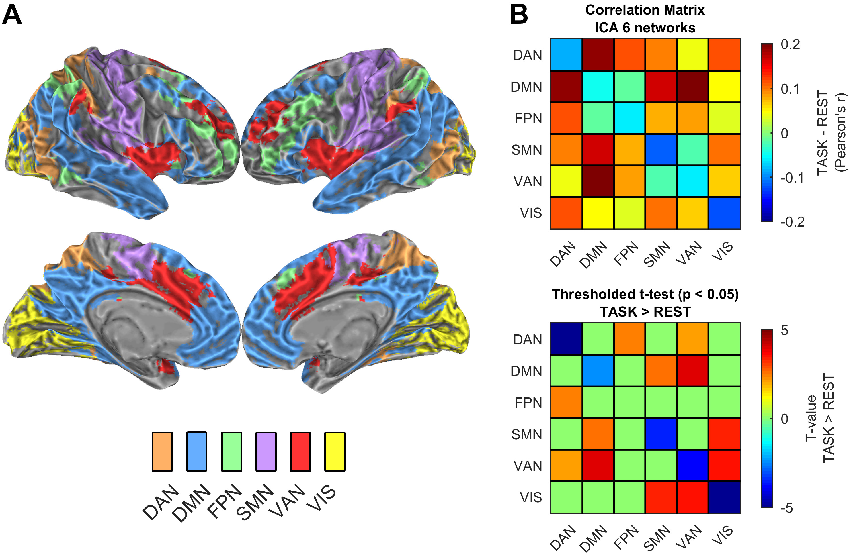
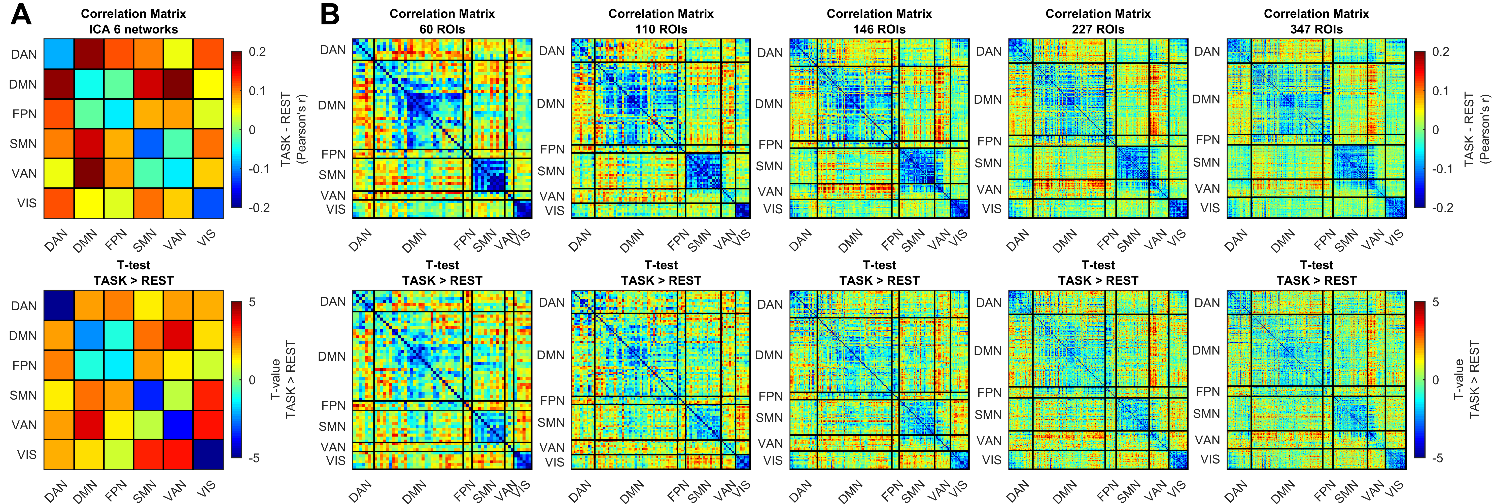
Six networks were identified by ICA decomposition4 of the first RS epoch (FIG1A). To probe the spatial scale at which FC changes happen, the cortex was also parceled into a variable number of ROIs, via a 2-levels analysis that built group level ROIs based on the similarity between each voxel time course5. ROIs were then classified into one of the ICA derived networks with a minimum overlap criterion (65%). ROIs with insufficient overlap were excluded, finally obtaining brain parcellations into 60, 146, 227, 347 and 477 ROIs. FC was estimated separately in each RS and WM epoch computing between-networks and between-ROIs correlations6. Within-network FC was also computed as the average of correlation values inside a given network. FC of homologous epochs were then averaged and their task-associated modulations were assessed via paired t-tests. Finally, correlations with WM performance was examined to assess the behavioral relevance of FC changes.
RESULTS
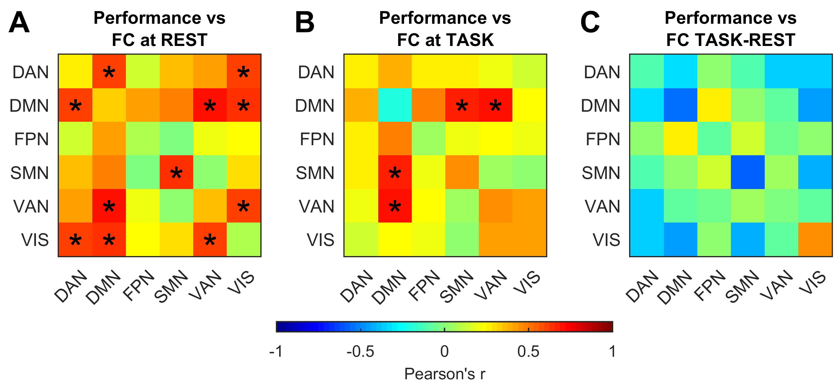
The WM task was associated with significant changes in FC, both within and between networks, but with opposite trends (FIG1B). Within-network FC showed significant reductions during task execution in all networks with the exception of FPN, while between-network FC showed increased FC in many network pairs. Although not all network pairs reached statistical significance, they all showed a tendency to increase FC during task (see unthresholded t-matrix, FIG2A). The same clear-cut pattern of decreased within and increased between-network FC was visible at each investigated spatial scale (FIG 2B). WM performance was positively correlated with FC at rest in several network pairs, which include the dorsal and ventral attention network and the default mode network (FIG3A). FC at task also showed positive correlations with performance (FIG3B), although with a smaller magnitude compared to rest. On the contrary, no significant correlation was found between performance and changes in FC (FIG3C).DISCUSSION
The opposite changes of between and within-network FC in response to sustained execution of the WM task depict a brain that reacts to stimuli by increasing cooperation among networks at the expense of reduced internal network coherence. From the largest scale (at the network level) to the smallest scale (477 ROIs) the pattern of FC changes was unchanged, indicating a scale-invariant feature of task-associated modulations. Despite such striking network behavior, performance did not show any clear-cut relationship with changes in FC, while performance was positively correlated with FC at rest and at task in agreement with previous studies7. The lack of linear correlation between performance and changes in FC suggests that the latter might be a simple epiphenomenon that does not carry any behavioral relevance. Alternatively, more complex non-linear relationships might underpin the behavioral correlates of the task-associated changes in FC. Further studies are warranted to shed light upon the matter.CONCLUSION
We further push the notion that the brain engagement in the external world goes beyond the well-studied localized task-evoked BOLD response, involving also a remarkable modulation of BOLD LFFs. The latter component cannot be overlooked if a comprehensive picture of the task-related brain activity is sought.Acknowledgements
No acknowledgement found.References
1. Damoiseaux, J. S., et al. "Consistent resting-state networks across healthy subjects." Proceedings of the national academy of sciences 103.37 (2006): 13848-13853
2. Cole, Michael W., et al. "Intrinsic and task-evoked network architectures of the human brain." Neuron 83.1 (2014): 238-251.
3. Behzadi, Yashar, et al. "A component based noise correction method (CompCor) for BOLD and perfusion based fMRI." Neuroimage 37.1 (2007): 90-101.
4. Beckmann, Christian F., and Stephen M. Smith. "Probabilistic independent component analysis for functional magnetic resonance imaging." IEEE transactions on medical imaging 23.2 (2004): 137-152
5. Craddock, R. Cameron, et al. "A whole brain fMRI atlas generated via spatially constrained spectral clustering." Human brain mapping 33.8 (2012): 1914-1928.
6. Biswal, Bharat, et al. "Functional connectivity in the motor cortex of resting human brain using echo‐planar mri." Magnetic resonance in medicine 34.4 (1995): 537-541
7. Hampson, Michelle, et al. "Brain connectivity related to working memory performance." Journal of Neuroscience 26.51 (2006): 13338-13343
Figures