2309
BOLD-fMRI comparison of olfactory responses in the mouse whole brain, with different odors and anesthesia1Faculty of Life Sciences, Kumamoto University, Kumamoto, Japan
Synopsis
Mice have well-developed olfactory systems, and the odor response throughout the mouse whole brain is an important target of olfactory research. We previously applied independent component analysis (ICA), which identifies periodically activated regions, to detect BOLD responses from odor-stimulated mice. In this study, we successfully discriminated olfactory responses from different odors, isoamyl acetate and musocone, in the mouse whole brain, using the BOLD-ICA method. In addition, we investigated the effects of urethane and medetomidine anesthetics on the olfactory responses. This study demonstrated the utility of the BOLD-ICA method to trace the real-time activation of the mouse whole brain.
Introduction
Olfaction is one of the essential perceptions for animals. Upon inhalation of odorant compounds, the olfactory bulb (OB) initially exhibits unique activation patterns depending on the kinds of odors. However, our understanding of the odor-stimulated activation patterns beyond the OB is still limited. To reveal how the brain discriminates different odors, the activation pattern over the whole brain must be investigated [1]. We are seeking to establish a method that can detect odor responses in the mouse whole brain and to reveal the mechanism of odor discrimination. The BOLD analysis is the method of choice to monitor real-time odor responses in rodents, which have well-developed olfactory systems. Some odor fMRI studies have been reported for the mouse OB [2], but the number of fMRI studies detecting the activation in the mouse whole brain is still limited [3]. One difficulty in investigating the whole brain by the BOLD method is that the mouse brain is smaller and thus the BOLD analysis is more likely to suffer from peripheral hemodynamic changes, making it harder to detect fMRI signals [4]. Thus, a robust method is needed to deal with the mouse whole brain. We previously reported a BOLD-fMRI method using repetitive odor stimulation and independent component analysis (ICA), which identifies periodically activated brain regions with high detectability [5]. The aim of this study is to discriminate the olfactory responses to different odors in the mouse whole brain and to investigate the effects of anesthetics on these responses, using the BOLD-ICA method.Methods
MRI experiments were performed with a 7.0 Tesla Bruker BioSpec 70/20 scanner and a mouse brain 2-channel phased array surface cryogenic coil (Bruker BioSpin). Mice (male C57BL/6, 8–10 weeks old) were anesthetized with urethane (i.p. 1.5 g/kg initial; 0.1 g/kg/hr supplemental) or medetomidine (i.p. 0.3 mg/kg initial; 0.1 mg/kg/hr supplemental). GRE-EPI images were acquired: TR/TE = 2000/21.4 ms; FOV = 1.28×1.28 cm2; matrix = 96×64; resolution = 200×200 µm2; slice thickness = 400 µm; number of slices = 20; NEX = 1; flip angle = 90°. At 1 min intervals, isoamyl acetate (IAA) or muscone vapor was mixed with the air flow and delivered to the mouse nose for 5 sec. We combined the data obtained from 8 mice, and analyzed them by group ICA with the FSL software. The MR signal transition components with the frequency of 16.6 mHz were selected, to identify the activated regions in the mouse brains. The peak intensity profiles were created by summing the intensity profiles of the 24 tasks over 8 mice.Results
Upon stimulations of IAA with a banana odor, brain responses in the piriform cortex (Pir) were detected by the BOLD-ICA method under urethane anesthesia (Fig. 1A). Upon stimulations with muscone, one of the natural musk compounds, brain responses in Pir were detected, but the time course of the responses was different from that in the case of IAA (Fig. 1B). Pir functions in the general olfactory pathway, and thus it is conceivable that Pir was activated by both IAA and muscone. The posterior lateral hypothalamus (PLH) was also activated by IAA (Fig. 1C), but not by muscone (Fig. 1D). Therefore, PLH is an odor-specifically activated region, in contrast to Pir. Next, we investigated the effects of anesthetics on the odor responses (Fig. 2). Under both urethane and medetomidine anesthesia, odor responses were evoked by IAA in Pir (Fig. 2A and B) and amygdala (Fig. 2C and D). However, the odor responses under medetomidine were larger than those under urethane.Discussion
By virtue of the elimination of noise signals by the BOLD-ICA method, the activation maps were simplified. Notably, the observed time course in response to the muscone-stimulation was different from the typical hemodynamic response (Fig. 1B). The conventional general linear model (GLM), which hypothesizes the typical hemodynamic response, fails to catch such responses, but the BOLD-ICA method actually could detect the response. The specific IAA-evoked activation of PLH (Fig. 1C) may indicate the activation of the feeding center, which is within PLH, by the banana-like odor of IAA. The larger odor responses under medetomidine anesthesia indicate that medetomidine mildly sedates the level of consciousness at this fixed dose, which is preferable for olfactory research.Conclusion
We successfully discriminated the olfactory responses from different odors in the mouse whole brain. This study demonstrated the utility of the BOLD-ICA method to trace the real-time activation of the mouse whole brain.Acknowledgements
No acknowledgement found.References
[1] Touhara, K. and Vosshall, L.B., Sensing odorants and pheromones with chemosensory receptors. Annu. Rev. Physiol. 2009; 71: 307–332
[2] Xu, F. et al., Odor maps of aldehydes and esters revealed by functional MRI in the glomerular layer of the mouse olfactory bulb. Proc. Natl. Acad. Sci. USA 2003; 100: 11029–11034
[3] Pain, F. et al., Visualizing odor representation in the brain: a review of imaging techniques for the mapping of sensory activity in the olfactory glomeruli. Cell. Mol. Life Sci. 2011; 68: 2689–2709
[4] Ielacqua, G.D. et al., Proc. Intl. Soc. Mag. Reson. Med. 2015; 23: 2037
[5] Funatsu et al. A BOLD analysis of the olfactory perception system in the mouse whole brain, using independent component analysis. Proc. Intl. Soc. Mag. Reson. Med. 2017; 25: 5363
Figures

Fig. 1 Comparison of olfactory responses to different odors in the mouse whole brain.
IAA-evoked responses in the piriform cortex (Pir) (A) and the posterior lateral hypothalamus (PLH) (C). Muscone-evoked responses in Pir (B) and PLH (D). The fMRI experiments were performed under urethane anesthesia. The ICA map and the signal time course of the activated region are shown in each panel. Yellow squares show the durations of the odor stimulations.
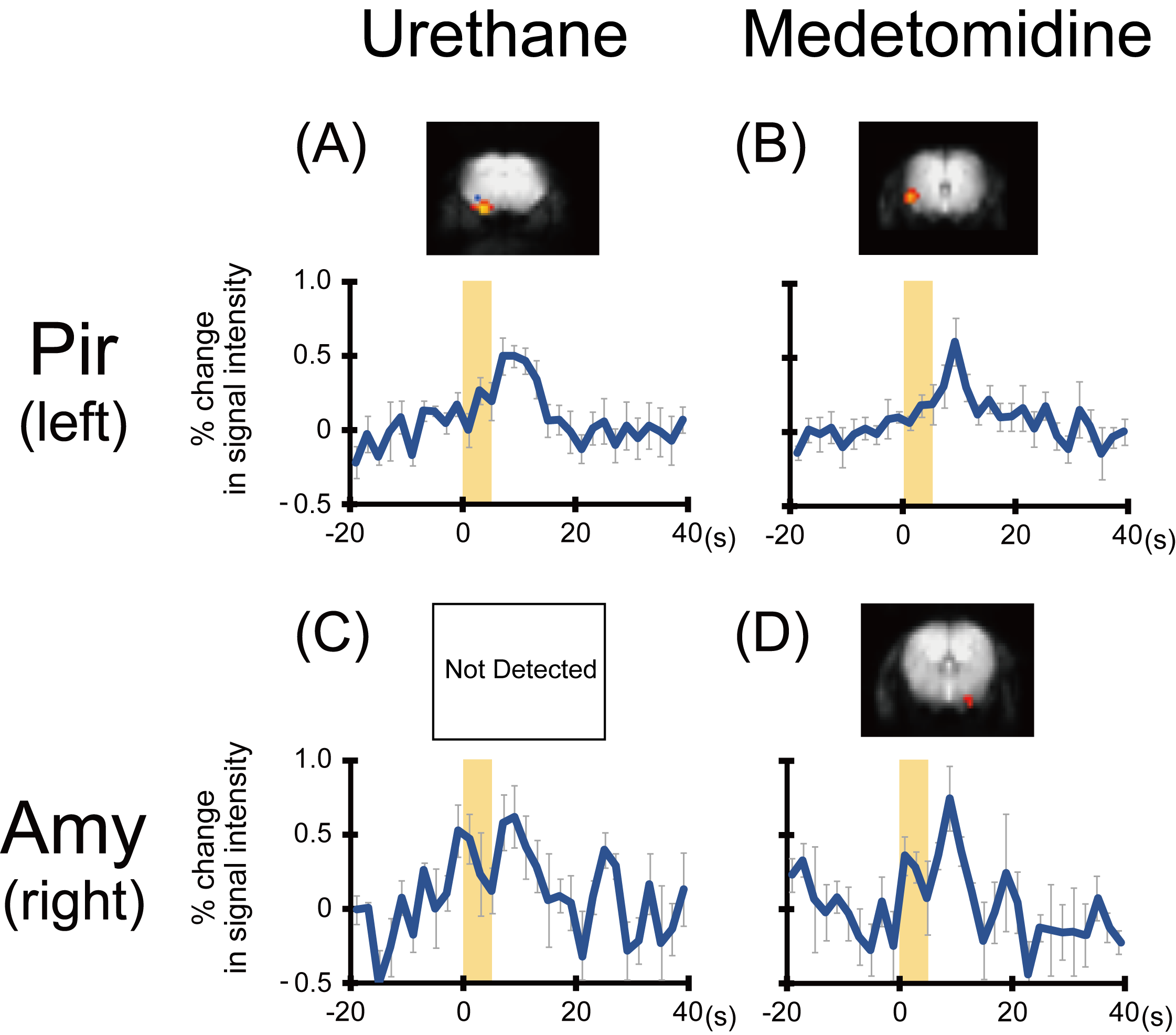
Fig. 2 Comparison of olfactory responses with different types of anesthesia in the mouse whole brain.
IAA-evoked responses in Pir under urethane (A) and medetomidine (B) anesthesia. IAA-evoked responses in amygdala (Amy) under urethane (C) and medetomidine (D) anesthesia. The ICA map and the signal time course of the activated region are shown in each panel. Yellow squares show the durations of the odor stimulations. The experimental data in Fig. 2A are the same as those in Fig. 1A, and shown for comparison.