2302
Comparison of neurovascular coupling and BOLD responses under medetomidine and isoflurane anesthesia in the rat somatosensory cortex.1Chiropractic, UQTR, Trois-Rivieres, QC, Canada, 2Anatomy, UQTR, Trois-Rivieres, QC, Canada, 3NeuroSpin, Commissariat à l’Energie Atomique-Saclay Center, Gif-sur-Yvette, France, 4Chiropractic, UQTR, Trois-Rivières, QC, Canada
Synopsis
In this study, we aimed at comparing the coupling between neuronal activity and hemodynamic changes evoked by hindpaw stimulation, under medetomidine and isoflurane anesthesia. Simultaneous recordings of local field potentials (LFP) and cerebral blood flow (CBF) were performed in the rat somatosensory cortex. In a separate experiment, hemodynamic changes evoked by hindpaw stimulation were measured using BOLD fMRI. The coupling between LFP amplitude and CBF changes was similar between isoflurane and medetomidine anesthesia. However, BOLD signal changes were smaller under isoflurane compared with medetomidine anesthesia. This suggests that isoflurane anesthesia may alter BOLD signal through alteration of O2-consumption or O2-saturation.
INTRODUCTION
Previous functional MRI studies revealed that the amplitude of blood oxygenation level dependent (BOLD) responses are smaller under isoflurane anesthesia compared with medetomidine anesthesia1, suggesting that these anesthetics influence neurovascular coupling differently. In this study, we aimed at investigating the mechanism of the different effects of isoflurane and medetomidine on BOLD responses. We hypothesized that the coupling between neuronal activity and vascular changes evoked by somatosensory stimulation would be different between anesthetics. We compared the amplitude of local field potentials (LFP) and cerebral blood flow (CBF) changes recorded simultaneously in the rat somatosensory cortex2-3, during electrical stimulation of the hindpaw (Experiment 1). In addition, we measured BOLD changes evoked by somatosensory stimulation under isoflurane and medetomidine anesthesia (Experiment 2), to confirm the reported difference between anesthetics.METHODS
Animals and anesthesia
Male Wistar rats (200 - 400 g) were used for Experiment 1 (n = 7) and Experiment 2 (n = 5). The respiration was controlled by mechanical ventilation and body temperature was maintained at 37 ± 0.5 °C. First, rats were anesthetized under isoflurane (1.5% with air). After the measurement of LFP and CBF or BOLD signal, isoflurane anesthesia was interrupted and replaced by medetomidine (0.1 mg/kg/h, i.v.). After stable recordings could be obtained, LFP and CBF or BOLD signal were measured again for comparison.
Electrical stimulation
In Experiment 1, the left sciatic nerve was stimulated for 10 s with 1-ms pulses at 5 Hz at an intensity ranging between 0.01 and 9.6 mA. In Experiment 2, electrical stimulation was applied for 30 s with subcutaneous needle electrodes inserted in the forepaw with 0.3-ms pulses at 7 Hz at an intensity of 2 mA.
Local field potentials
Local field potentials were measured with a microelectrode at a 600 µm depth in the hindpaw representation of the primary somatosensory cortex (1-300 Hz), sampled at 5 kHz and recorded for offline analyses. LFP amplitude was measured as the peak-to-peak amplitude from stimulus onset to 50 ms poststimulus.
Cerebral blood flow
A Laser-Doppler probe was placed on the cortical surface, as close as possible to the microelectrode without achieving contact. CBF was measured with a time constant of 3 s, sampled at 100 Hz and recorded for offline analyses. CBF changes were measured as the onset-to-peak amplitude.
Neurovascular coupling
Neurovascular coupling was calculated as the ratio of CBF changes on LFP amplitude using stadardized values (T-scores).
Magnetic Resonance Imaging
FMRI acquisition was conducted with Bruker 7T scanner using a 4-ch rat brain array coil. FMRI images were acquired using a gradient-echo EPI sequence, TR/TE = 2,000/11 ms, spatial resolution = 200 x 200 x 800 µm3, 8 slices, for 5.5 min (165 volumes). During measurements, respiration was maintained at a rate of 50/min and body temperature at 37 ± 0.5 °C. Anatomical images were acquired for spatial correction using multi-slice rapid acquisition with relaxation enhancement (RARE), using the same FOV and resolution as for EPI: TEeff/TR = 36/2500 ms, RARE factor = 8.
Image processing of fMRI data
Slice timing correction and spatial correction of images were performed with SPM8 (Welcome Trust Center for Neuroimaging, UK). The time-course in was extracted in the forepaw representation of the primary somatosensory cortex using a script written in Matlab (Mathworks, MA).
RESULTS
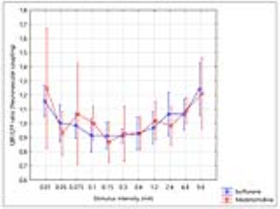
LFP amplitude and CBF changes were similarly coupled under isoflurane compared with medetomodine anesthesia for all intensities (interaction: F(10,60)=0.4, p=0.9; see Fig. 1). However, Student T-test revealed that the amplitude of BOLD signal changes under isoflurane anesthesia was significantly smaller compared with medetomidine anesthesia (p<0.05; Fig. 2).DISCUSSION
The novel finding of this study is that neurovascular coupling (CBF/LFP ratio) measured in the primary somatosensory cortex was not significantly different between isoflurane and medetomidine anesthesia during hindpaw stimulation. In contrast, the amplitude of BOLD responses under isoflurane anesthesia was smaller compared with medetomidine anesthesia. These results suggest that isoflurane could affect O2-consumption or O2-saturation during somatosensory stimulation, in spite of comparable neuronal activation.CONCLUSION
Future studies are needed to confirm that altered O2-consumption or O2-saturation causes different BOLD responses between isoflurane and medetomidine anesthesia without affecting neurovascular coupling.Acknowledgements
This study was funded by the Natural Science and Engineering Research Council (NSERC) of Canada.References
[1] Chao TH, Chen JH, Yen CT. Repeated BOLD-fMRI imaging of deep brain stimulation responses in rats. PLoS One. 2014;9(5):e97305.
[2] Jeffrey-Gauthier, R., J.P. Guillemot, and M. Piche, Neurovascular coupling during nociceptive processing in the primary somatosensory cortex of the rat. Pain, 2013. 154(8): p. 1434-41.
[3] Uchida, S., S. Bois, J.P. Guillemot, H. Leblond, and M. Piche, Systemic blood pressure alters cortical blood flow and neurovascular coupling during nociceptive processing in the primary somatosensory cortex of the rat. Neuroscience, 2016. 343: p. 250-259.
Figures

