2291
MANGANESE ENHANCED MRI: A METHOD IN ORDER TO VALIDATE PHYSILOGICAL MARKERS OF TINNITUS IN RODENTS1Charles Coulomb Laboratory, Montpellier, France, Metropolitan, 2CILCARE, Montferrier sur Lez, France, Metropolitan, 3Laboratoire de Neurosciences intégratives et Adaptatives, Marseille, France, Metropolitan
Synopsis
The present study is designed to show physiological markers of tinnitus in rodents. The tinnitus is an auditory phantom sensation experienced in absence of an external stimulus. The prevalence of tinnitus shows a worrying growth curve with the development of new lifestyles (exposure to noise, urbanization, …). One promising tool is used, called Manganese Enhanced MRI (MEMRI). The use of manganese chloride as an MRI contrast agent enables to follow brain neuronal activity. T1-weighted MRI images are collected in order to investigate the specific areas activated in presence of tinnitus or not. Two analysis methods are used: Statistical analysis by Signal to Noise Ratio (SNR) and T2 rate cartography. Results enable to shown a complementary between the two analysis methods and allows us to discriminate between healthy and tinnitus rats.
INTRODUCTION
Tinnitus is an auditory phantom sensation (crackling, humming, whistling) experienced in absence of an external stimulus. This common disorder affects 10-15% of the population at one time or another in life. It creates discomfort sometimes associated with pain. The prevalence of tinnitus shows a worrying growth curve. The new lifestyles of developed and emerging countries (exposure to noise, urbanization, …) accelerate this trend and turn it a major public health problem.METHODS
The present study is designed to show physiological markers of tinnitus in rodents. One promising Magnetic Resonance Imaging (MRI) tool is used, called Manganese Enhanced MRI (MEMRI). The use of manganese chloride as an MRI contrast agent enables to follow brain neuronal activity. In fact, paramagnetic manganese ions (Mn2+) accumulate in active neurons through voltage-gated calcium channels. The protocol based on MEMRI technic uses an intratympanic injection on rats with the salicylate model of tinnitus induction. Moreover, T1-weighted MRI images are collected in order to investigate the specific areas activated in presence of tinnitus or not. Two analysis methods are used: Statistical analysis by Signal to Noise Ratio (SNR) and T2 rate cartography.RESULTS
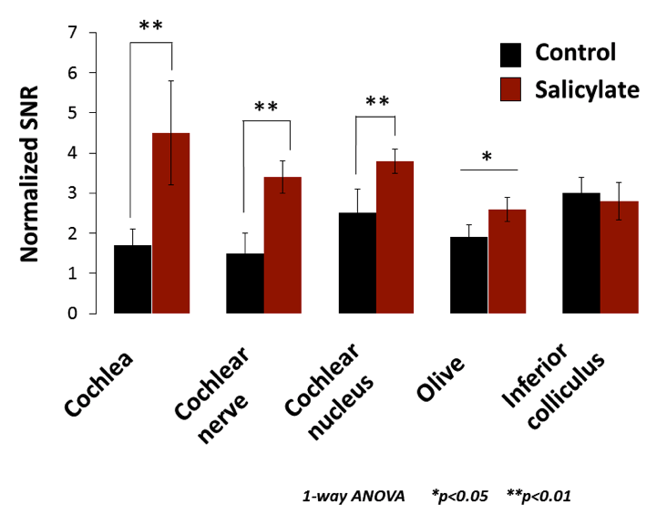
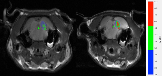
Statistical analysis reveals that a SNR enhancement is observed in the main auditory areas (cochlea, cochlear nerve, cochlear nucleus, superior olivary complex) specifically in disabled rats. To extend these results, a new cartography was developed in this project using homebuilt program developed under Matlab interface. T2 rate cartography is based on high relaxation rates shown in tissues containing MnCl2. The aim of this tool is to detect precisely the accumulation of MnCl2. The recent works enabled to segment a particular auditory area, the contralateral inferior colliculus and to apply it to the T2 rate cartography.
DISCUSSION
Statistical analysis by SNR and T2 rate cartography are two complementary analysis methods. In fact, T2 rate cartography allows to local auditory regions with high concentration of MnCl2, and provide accurate quantitative data enables us to discriminate between heathy and tinnitus rats.CONCLUSION
The development and quantification of T2 rate cartography open a new axis in tinnitus researches with MEMRI method. Our innovative study presents some potential applications in auditory research.Acknowledgements
No acknowledgement found.References
1. Adjamian P, Hall DA, Palmer AR, Allan TW, Langers DR. Neuroanatomical abnormalities in chronic tinnitus in the human brain. Neurosci Biobehav Rev. 2014, 45:119-33.
2. Cacace AT1, Brozoski T2, Berkowitz B3, Bauer C2, Odintsov B4, Bergkvist M5, Castracane J5, Zhang J6, Holt AG7. Manganese enhanced magnetic resonance imaging (MEMRI): a powerful new imaging method to study tinnitus. Hear Res. 2014 311:49-62.
3. Cazals Y. Auditory sensori-neural alterations induced by salicylate. Prog Neurobiol. 2000 Dec;62(6):583-631. Review. 4. Cazals Y, Horner KC, Huang ZW. Alterations in average spectrum of cochleoneural activity by long-term salicylate treatment in the guinea pig: a plausible index of tinnitus. J Neurophysiol. 1998 Oct;80(4):2113-20.
Figures