2279
MR Elastography in a mouse model of Alzheimer’s disease: 5XFAD Mice1Department of Bioengineering, University of Illinois at Chicago, Chicago, IL, United States, 2Department of Anatomy and Cell Biology, University of Illinois at Chicago, Chicago, IL, United States
Synopsis
In vivo magnetic resonance elastography (MRE) experiments on the 5XFAD Alzheimer’s disease (AD) mouse model were conducted. The AD and Control mice were in the age group of ~1 month (n = 2 for both) and 3~4 months (n = 5 for both). Median stiffness values were measured over different regions of the brain. The overall brain tissue was stiffer in the disease model when compared to the control, with results being significant at the 3~4-month time point. Further experiments are underway at the 1-month time-point for conclusive age-based comparisons.
Target Audience
Elastography, Alzheimer’s disease, and neurodegenerative disease diagnosis.Purpose
Through this study, we are aiming to establish the early diagnostic potential of MRE in assessing Alzheimer’s disease (AD). AD is known to be accompanied by extensive Amyloid-β (Aβ) plaque deposition. In our initial phase of the study, we opted to use the APPswe/PS1dE9 transgenic mice model which carry the transgene for both the human amyloid precursor protein (APP) and the presenilin 1 (PS1) protein and exhibit extensive Aβ deposits at 6 months of age.1 The preliminary study findings were presented at the 2017 Joint Annual Meeting ISMRM-ESMRMB.2 As gathering data from such a mouse model at different time-points presents a serious logistical challenge, in this current study, we used a new animal model i.e. 5XFAD mice. This way, we are still using a model which carries similar transgenes as the APPswe/PS1dE9 mice, but at the same time this is a more aggressive model in terms of disease expression, showing plaque deposits as early as 2 months of age.1Methods
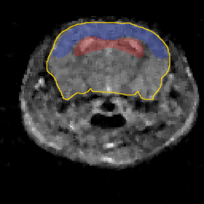
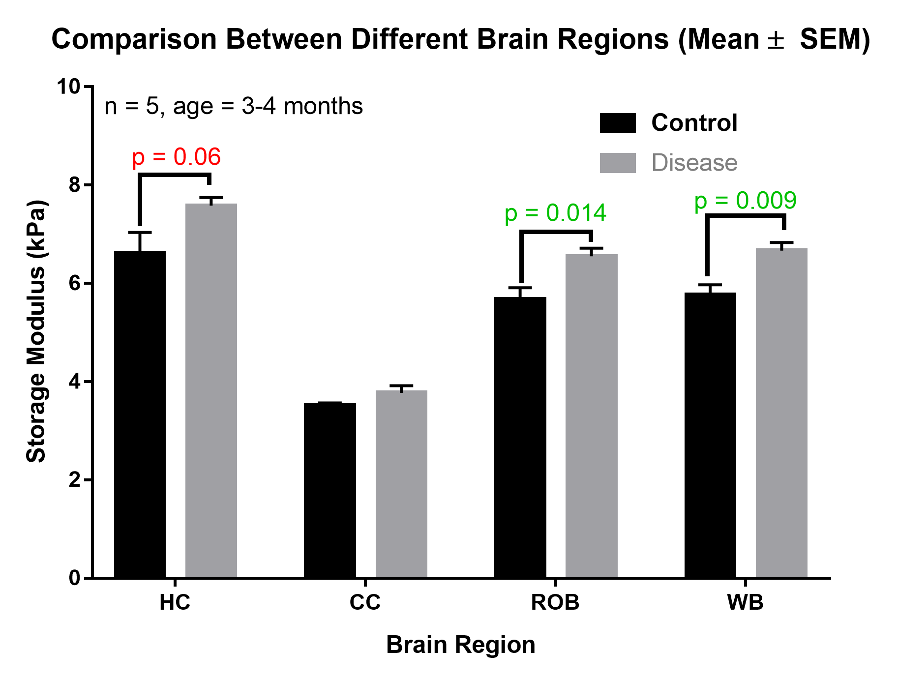
Data was gathered from 2 groups of female, Bax:5XFAD mice (Disease) and Bax:Nestin mice (Control), with one group being at ~1 month (n = 2) of age and the other at 3~4 months (n = 5). A 9.4T pre-clinical MRI scanner at the Research Resources Center of the University of Illinois at Chicago was used to perform the measurements. In the elastography experiments, a vibrating bite bar type actuator induces mechanical shear waves in the mouse brain. Experimental details and SLIM-MRE3 imaging parameters are as follows: 38 mm volume radiofrequency (RF) coil, 16 mm field of view axial slices, 250 μm isotropic voxel size, TE/TR = 16.24/1000 ms, 3 averages, 1000 Hz actuation frequency, 250 mT/m motion encoding gradient (MEG) strength, 10 MEG cycles, and 8 time steps, with a total scan time of 51 min. 12 secs. Using the curl operator on noise-filtered complex wave images, the complex shear modulus was calculated by the algebraic inversion of the Helmholtz equation. Storage modulus values (real part of the complex modulus) over four region of interests (ROIs) were calculated. ROI-1 is the overall area of the mouse brain (WB) as seen in figure 1 (marked with yellow border). ROIs-2&3 (blue and red highlighted region in figure 1) of the hippocampal area (HC) and the cerebral cortex (CC) were manually segmented from MRI magnitude images. Finally, the rest of the brain (ROB) corresponds to ROI-1 without ROI-2 and ROI-3 and is denoted with ROI-4.Results
Based on the comparison of overall brain stiffness (storage modulus), we measured an increase in brain stiffness in the disease model as compared to the control at both age groups. The difference was highly significant (p-value < 0.01) in case of the group at 3~4 months, while current limitations of sample size at 1 month did not reveal a significant difference. For the older age group we also observed close to significant differences in the hippocampal region. In general, the stiffness across most brain regions was higher for the disease model (5XFAD) as compared to the control mice.Discussion
Further experiments will be performed in the near future to get additional mice data at both age groups, so that a more comprehensive comparison can be performed on the effects of disease progression on brain stiffness.Acknowledgements
The experimental costs were funded by the UIC Chancellor’s Discovery Fund for Multidisciplinary Pilot Research, granted to Dr. Klatt (Dept. of Bioengineering) and Dr. Orly Lazarov (Dept. of Anatomy and Cell Biology).References
1. Lee, J.-E. & Han, P.-L. An Update of Animal Models of Alzheimer Disease with a Reevaluation of Plaque Depositions. Exp. Neurobiol. 22, 84 (2013).
2. Majumdar, S. & Klatt, D. In vivo cerebral MR elastography in a mouse model of Alzheimer ’ s disease : preliminary results. Joint Annual Meeting ISMRM-ESMRMB Available at: http://cds.ismrm.org/protected/17MPresentations/abstracts/1375.html.
3. Klatt, D., Yasar, T. K., Royston, T. J. & Magin, R. L. Sample interval modulation for the simultaneous acquisition of displacement vector data in magnetic resonance elastography: theory and application. Phys. Med. Biol. 58, 8663–75 (2013).
Figures