2236
A Parallel Scheme of RF Irradiation and Data Acquisition for Chemical Exchange Saturation Transfer (CEST) MRI1Korea Advanced Institute of Science and Technology (KAIST), Daejeon, Republic of Korea
Synopsis
The chemical exchange saturation transfer (CEST) MRI usually requires long RF irradiation before every data acquisition to achieve the steady-state CEST mechanism. To eliminate the repeatedly required RF irradiations and to increase the scan efficiency, a new CEST MRI technique that performs the RF irradiation in parallel with data acquisition is developed. The results of MR experiments demonstrate the feasibility of the proposed technique in amide proton CEST.
Introduction
Chemical exchange saturation transfer (CEST) MRI is an effective method to detect dilute labile protons.1 In conventional CEST imaging, the long preparation time of RF irradiation, which employs a continuous long RF pulse or repetitive RF pulses, is required before a data acquisition to attain the steady-state CEST mechanism. Since the steady-state CEST is disturbed by the imaging data acquisition, the RF irradiations should be repeated for every data acquisition. Consequently, these long RF irradiations prolong imaging time. In this abstract, we propose a new CEST MRI technique that perform the RF irradiation in parallel with the data acquisition, so that the steady-state CEST effect can be maintained during data acquisitions and the scan efficiency can increase.Methods
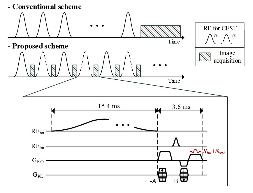
The proposed scheme for CEST MRI is based on the irradiation scheme of repetitive RF pulses with time intervals, so called pulsed-CEST preparation.2 Fig. 1 shows the conceptual pulse sequence of the proposed scheme compared with the conventional pulsed-CEST. The proposed scheme acquires image signals at the time interval between the CEST RF pulses, which is intended for low specific absorption rate (SAR) in the pulsed-CEST preparation. The pulse sequence for acquiring image signals is the steady-state free precession (SSFP), of which gradients also function as spoiler gradients for pulsed-CEST preparation. The steady-state condition of both CEST mechanism and image signals are maintained simultaneously during entire scanning. Once the steady-state conditions are achieved, there is no need to perform other CEST preparations in acquiring additional image signals. Therefore, the proposed method could increase the efficiency of scan time in CEST MRI.
Although the signals (Scest) excited by the RF pulses of CEST preparation are not spoiled perfectly and are combined with image signals (Sim) in data acquisition, we can eliminate Scest from the acquired signals (Sacq) in the spatial domain due to the property of acquired Scest.3 From the proposed imaging sequence, Sacq are represented as follows:
$$S_{acq}(k_{x},k_{y})=S_{im}(k_{x},k_{y})+S_{cest}(k_{x}+c_{1},c_{2})e^{j\pi k_{y}}$$
where kx and ky are the readout and phase-encoding dimensions of k-space respectively, and c1 and c2 are constants induced by the gradients of the pulse sequence for image signals. $$$e^{j\pi k_{y}}$$$ is induced by the alternating RF phases of the CEST preparation (Fig. 1). All echoes of Scest have identical phase accumulation (c2) in the phase-encoding dimension. The acquired signals are then 2D inverse Fourier transformed ($$$F_2^{-1}$$$) as follows:
$$I_{acq}=F_2^{-1}[S_{acq}(k_{x},k_{y})]$$
$$=I_{im}(n_{x},n_{y})+P_{cest}(n_{x})\cdot\delta(n_{y}-\frac{N_{y}}{2}),$$
$$P_{cest}(n_{x})=\sum_{n_{y}}I_{cest}(n_{x},n_{y})e^{j2\pi (c_{1}n_{x}/N_{x}+c_{2}n_{y}/N_{y})}$$
where Iim and Icest are the spatial information of Sim and Scest, respectively. Ny is the number of phase-encodings. Since all echoes of Scest are identical in the ky dimension of Sacq, they can be Fourier-transformed into a line component ($$$P_{cest}(n_{x})$$$) in Iacq, which is located on the edge of the field of view (FOV). Therefore, the signals from the CEST RF pulses are spatially separated from image signals, so that they can be eliminated by a simple mask operation excluding the edge of FOV.
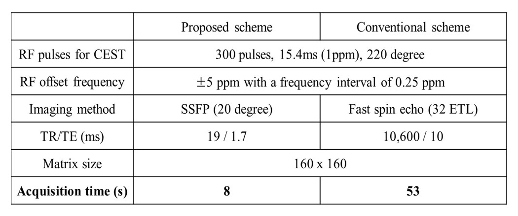
MR experiments were performed with a KAIST 3.0T MRI system (Siemens MAGNETOM Verio, Erlangen, Germany) to demonstrate whether the proposed scheme could provide CEST-contrast images similar to those from the conventional pulsed-CEST. Fresh white egg phantom was used to visualize the amide proton CEST at 3.5 ppm.4 The parameters used for two methods are summarized in Table 1.
Results
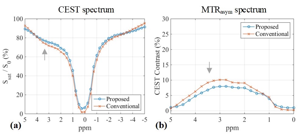
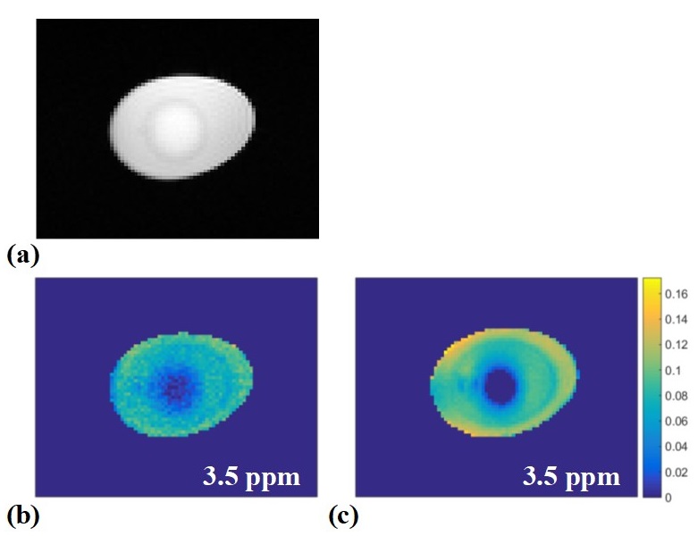
CEST spectra measured at fresh white egg are shown in Fig. 2(a), and the magnetization transfer ratio (MTR) asymmetry were analyzed as shown in Fig. 2(b). The signal decay induced by CEST effect appears around 3.5 ppm, which is the frequency of amide protons. The resultant CEST images at 3.5 ppm are shown in Fig. 3. Although the CEST contrast of the proposed scheme is 2 % lower than that of the conventional scheme, the time efficiency of the proposed scheme is about 7 times higher than that of the conventional scheme.Discussion & Conclusion
The proposed scheme is a fast imaging technique for CEST MRI, which maintains the steady-state CEST effect and simultaneously acquires the image data. The CEST contrast of the proposed scheme is comparable to that of the conventional scheme, even though the water protons are excited during the CEST preparation. MR experiments with egg phantom demonstrate that the proposed CEST scheme could visualize amide proton transfer within a few seconds.Acknowledgements
This research was supported by the Brain Research Program through the National Research Foundation of Korea (NRF) funded by the Ministry of Science, ICT & Future Planning (2014M3C7033999).
This research was supported by a grant of the Korea Health Technology R&D Project through the Korea Health Industry Development Institute (KHIDI), funded by the Ministry of Health & Welfare, Republic of Korea (grant number : HI14C1135)
References
1. van Zijl P, Zhou J, Mori N, et al. Mechanism of magnetization transfer during on‐resonance water saturation. A new approach to detect mobile proteins, peptides, and lipids. Magn Reson Med. 2003;49(3):440-9.
2. Sun PZ, Benner T, Kumar A, et al. Investigation of optimizing and translating pH‐sensitive pulsed‐chemical exchange saturation transfer (CEST) imaging to a 3T clinical scanner. Magn Reson Med. 2008;60(4):834-41.
3. Kim B, Seo H, Park H, et al. A Retrospective Cardiac Gating Method using Simultaneously Acquired Navigator. In Proceedings of the 25th Annual Meeting of ISMRM, Hawaii, 2017. p. 1282.
4. Lu J, Zhou J, Cai C, et al. Observation of true and pseudo NOE signals using CEST‐MRI and CEST‐MRS sequences with and without lipid suppression. Magn Reson Med. 2015;73(4):1615-22.
Figures