2235
Feasibility of ACIDOCEST using Iodixanol in a Rat Glioma Model1Radiology, University of Pennsylvania, PHILADELPHIA, PA, United States
Synopsis
In vivo pH mapping within tumor and kidney have been successfully demonstrated in both preclinical and clinical settings using MRI based imaging modality, known as AcidoCEST. This method uses iodinated contrast agents (ICAs) as exogenous contrast agent which are normally used in CT scans. So far, these methods have mainly utilized CEST contrast from to the amide peaks (~4.2, 5.6 ppm) ICAs. We demonstrate the feasibility of detecting the CEST contrast from both hydroxyl groups (~0.8 ppm) and amide groups (~4.2 ppm) from Iodixanol in the glioma model.
INTRODUCTION
In vivo pH mapping within tumor and kidney have been successfully demonstrated in both preclinical (1,2) and clinical (3) settings using MRI based imaging modality, known as AcidoCEST. Given the abundance of exchanging protons present in hydroxyl and amide groups, iodinated contrast agents (ICAs), approved for clinical CT diagnoses, have been used as exogenous AcidoCEST contrast agents. Most of ICAs have amide protons in two distinct chemical environments, making CEST contributions at ~4.2 ppm and ~5.6 ppm and a common approach is to use their relative CEST contributions to make pH mapping independent of the concentrations. These peaks are neither contaminated by the direct saturation, nor by CEST contributions from other endogenous metabolites which mostly contributes to < 4 ppm. However, the contribution of 5.6 ppm peak vanishes at higher pH (~6.8-7.4) and it is difficult to perform the ratiometric analysis in this pH range. We demonstrate for the first time the feasibility of detecting CEST contributions from hydroxyl peaks and amide peaks of Iodixanol in the glioma model (9L Model).METHODS
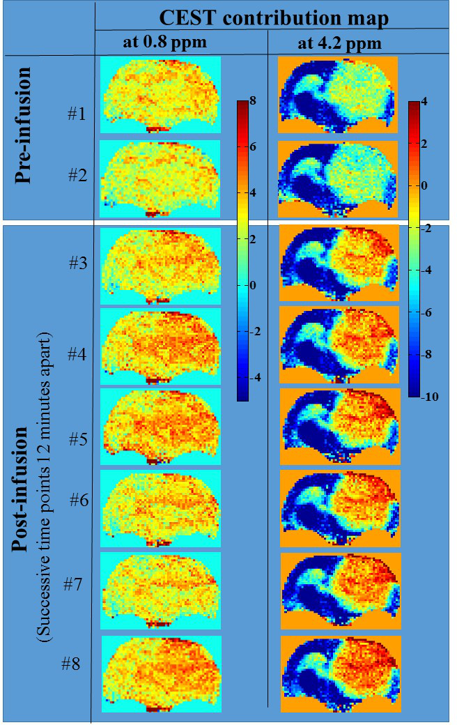
Phantom Experiment: High resolution NMR spectra of Iodixanol (Sigma-Aldrich, USA) solution (prepared at 100 mM, pH 7) were acquired using 400 MHz vertical bore scanner (Bruker, Germany) at three temperatures: 37ºC, 22ºC and 7ºC, employing the single pulse-acquired spectroscopy with TR=4s, #averages=32, with no water suppression. Solution phantoms with varying pH (pH = 6.0-6.8, concentration= 20 mM; 37 °C), were used to evaluate the CEST properties of Iodixanol at 9.4T (Agilent, USA), with sequence parameters identical to those used in animal experiment. Animal Experiment: A Wistar rat was injected with 9L glioma cells intracranially 3 weeks prior to the experiment. In vivo CEST MRI was performed at 9.4T MRI scanner, with rat being maintained under 1-1.5% isoflurane in O2 (supply: 1 L/min) and external heat source was used to maintain body temperature at (37±0.5)oC. CEST Sequence: The parameters were: slice thickness=4 mm, GRE flip angle= 15º, GRE readout TR=5.6ms, TE=2.7ms, FOV=30×30mm2, matrix size=128×128, T1 delay=6s, saturation duration 2s, saturation power 2.35 µT. CEST images from 0 to ±7.0 ppm were collected with 0.25 ppm step-size. B0 correction was performed by acquiring WASSR images at 0.23 μT from -1 to +1 ppm with 0.1 ppm step-size, with other parameters identical to CEST. Asymmetry analyses were performed at two offsets: 0.8 ppm, 4.4 ppm. Following the acquisition of the baseline CEST, Iodixanol was administered via tail vein. First a bolus dosage of 1000 µl lasting 2 minutes was administrated, followed by the constant infusion at the rate of 1000 µl/hour. Post-infusion scans were started 5 minute post-bolus, with the time resolution of ~12 minutes.RESULTS & DISCUSSIONS
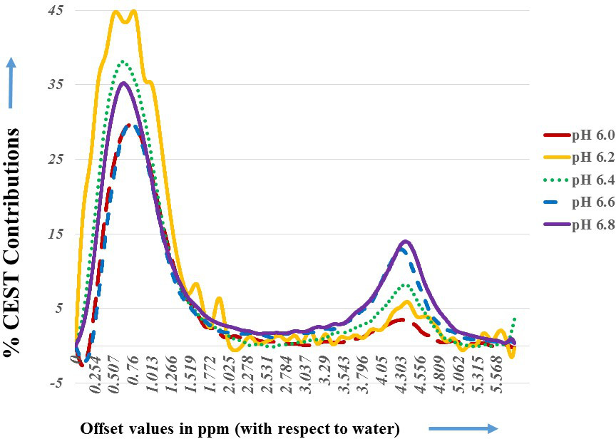
The high-resolution 1H-NMR spectra of Iodixanol showed the sharp resonance from amide proton at 4.4 ppm (Fig. 1) downfield of water at (7oC, 22oC and 37oC). On the other hand, the resonance from the –OH appeared as a broad shoulder (~1 ppm downfield of water) only at the lower temperature of 7oC. The absence of this shoulder at higher temperature (22oC and 37oC) could be attributed to the fast exchange of –OH protons. The Iodixanol phantom study showed a broad CEST peak at ~0.8 ppm and 4.4 ppm (Fig. 2) for the pH-value (6.0-7.0). The ratio of CEST-contrasts at 4.4 ppm peak to 0.8 ppm peak was confirmed to be linearly correlated with pH. For the CEST experiment on glioma model, elevated CEST contrasts were observed for both offset values (2-3% for 0.8 ppm, 3-5% for 4.4 ppm) post-infusion (Fig. 3). Additional studies with more rat tumors will be performed in the near future to assess the accuracy of pH measurements with Iodixanol.Acknowledgements
This project was supported by the National Institute of Biomedical Imaging and Bioengineering of the National Institutes of Health through Grant Number P41-EB015893.References
1.Moon B. F. et al., Contrast Media Mol Imag. 2015 November ; 10(6): 446–455. DOI:10.1002/cmmi.1647
2. Longo, Dario L., et al., J. of the Am. Chem. Society 136 (41): 14333-14336. doi:10.1021/ja5059313
3. Jones, K. M., et al., Mol Imaging Biol (2016), DOI: 10.1007/s11307-016-1029-7
Figures