2233
Optimization of OH-CEST contrast at 3T for clinical application of glucoCEST MRI1High-Field Magnetic Resonance, Max Planck Institute for Biological Cybernetics, Tuebingen, Germany, 2Istituto di Biostrutture e Bioimmagini, Consiglio Nazionale delle Ricerche (CNR), Torino, Italy, 3Dipartimento di Biotecnologie Molecolari e Scienze per la Salute, Università degli Studi di Torino, Torino, Italy, 4Diagnostic & Interventional Neuroradiology, University Hospital Tuebingen, Tuebingen, Germany
Synopsis
A 3D snapshot CEST sequence is optimized for contrast originating from hydroxyl groups of glucose molecules. Multi-B1-multi-pH measurements allow fitting of exchange rates of four glucose hydroxyl groups, which are then used to optimize pre-saturation parameters in simulation. The optimal protocol gave highly reproducible signals in 6 healthy volunteers, and showed no contrast when tested in a brain tumor patient. This protocol provides a robust baseline for glucose injection studies.
Purpose
Gluco-CEST and –CESL might go clinical soon1–4. To improve glucoCEST protocols we aim to measure glucose QUESP for various pH and temperature to get a valid glucose hydroxyl exchange rate estimation under physiological conditions by (i) simultaneous B1-pH Bloch-McConnell fitting, (ii) using this information for numerical optimization of GlucoCEST/CESL preparation at clinical field strengths, and (iii) applying this method for intrinsic OH-CEST measurement in a reproducibility study at 3T.Methods
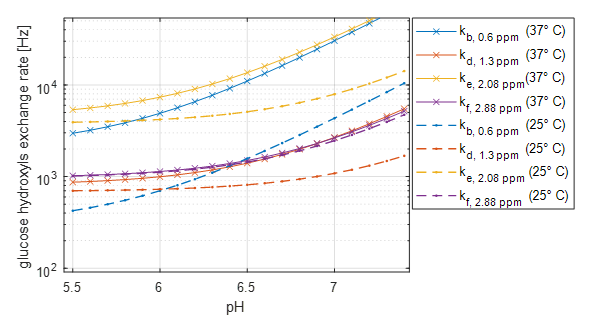
Seven tubes of 20 mM glucose solution in 1x PBS with different pH were measured at 2 temperatures for 6 different B1 levels between 0.5 and 3µT for 5s at B0=7T (Bruker). This stack of 7x6=42 Z-spectra (normalized and B0 corrected) was fitted simultaneously by a multi-Bloch-McConnell fit including the pH dependence for the exchange rate of each –OH pool x= b,d,e,f
$$k_x = k_{x1} \cdot10^{pH-7} + k_{x0}$$
The chemical shifts were assumed to be fixed at be δb=0.66 ppm, δd=1.28 ppm, δe=2.08 ppm and δf=2.88 ppm. We further assumed the concentration fraction of the –OH groups reflect their effective proton number of b:1, d:3, e:0.37 and f:(1-0.37) leading to the relative fractions at 20mM: 1.8018e-04, 5.4054e-04, 0.37·1.8018e-04 1.8018e-04, and (1-0.37) ·1.8018e-04, respectively. The R1 values for the water pool for both temperatures were provided to the fit.
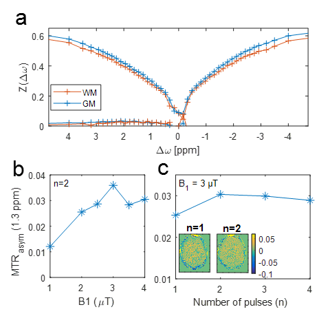
In vivo measurement with a presaturated 3D-GRE snapshot sequence5 was performed in 6 healthy volunteers in a Siemens PRISMA 3T scanner after providing written informed consent. Motion and B0 corrected Z-spectra (37 offsets between -3 and 3ppm) were evaluated by MTR asymmetry analysis.
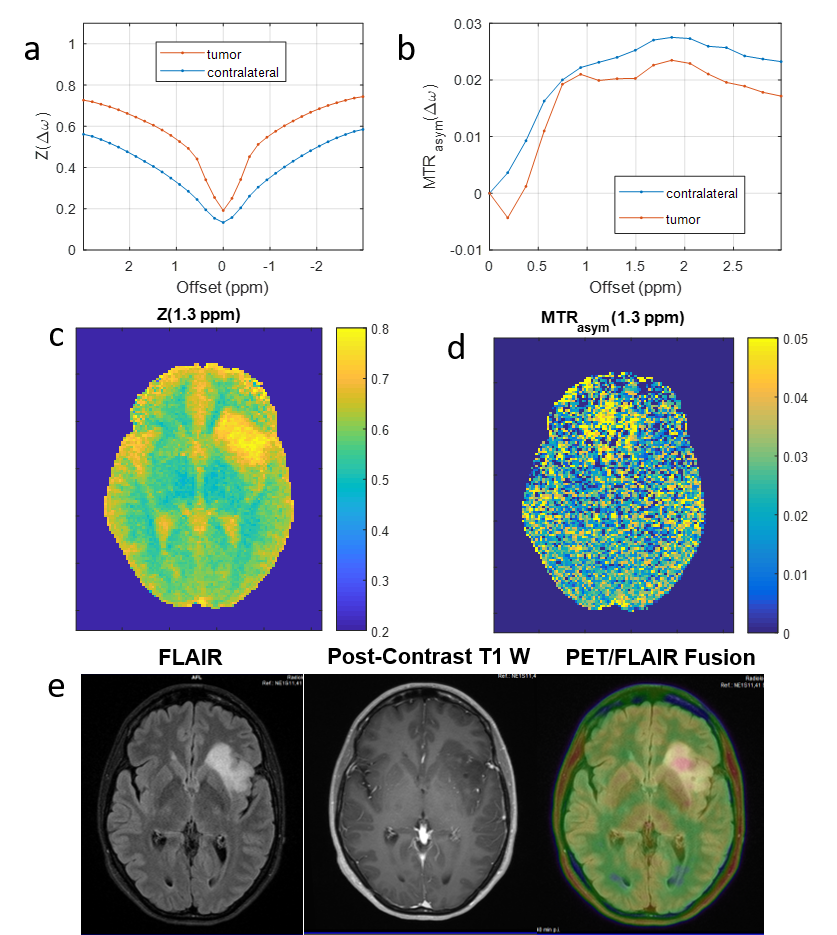
After validation for reproducibility, the OH-CEST MRI was included in the local routine PET/MR protocol. Clinical validation of the protocol was performed in one patient (scanned with Siemens Verio PET/MR) with suspected low-grade glioma.
Results
The Bloch-McConnell (BM) fit for the multi-pH-multi data fitted at 37° yields exchange rates of 1200 Hz, 2997 Hz, 3543 Hz, and 5863 Hz at pH=7.2 T=37° (Figure 1). Using these values in Bloch-McConnell simulations reveals that when taking into account direct water saturation and MT, the optimal pre-saturation for glucoCEST contrast in terms of CNR in grey matter at 3T is two 100ms pulses of B1 = 3µT at 1 ppm after long recovery (data not shown).
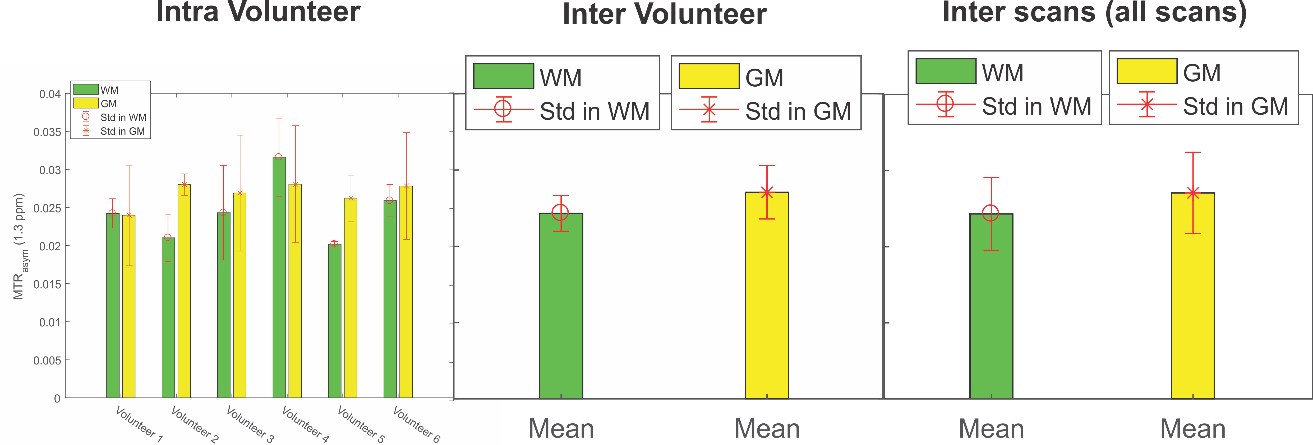
This presaturation was applied for intrinsic OH-CEST MRI of 6 healthy volunteers which showed a positive MTR asymmetry at 1.3 ppm with similar results for optimal B1 and number of pulses (Figure 2). MTRasym was highly reproducible with average values of 2.7% in GM and 2.4% in WM, and inter scan fluctuation <0.5% (Figure 3).
After this reproducibility validation, the OH-CEST MRI was applied in a brain tumor patient - still without glucose injection - with similar outcome in contralateral tissue (Figure 4). Interestingly, although the tumor is clearly visible in the Z-value image at 1.3 ppm (Figure 4c) due to high fluid content, the MTRasym looks very homogeneous (Figure 4d).
Discussion
The optimized OH-CEST protocol yields reliable and flat contrast in the human brain. First application in tumors without injection also revealed that the OH-CEST signal in tumor tissue stays similar to contralateral tissue signals (Figure 4). Thus, the proposed OH-CEST protocol forms a flat baseline for any injection experiments in tumor patients.
The relatively short but strong irradiation is especially selective for intermediate to fast exchanging protons. Thus amides, NOE and semi-solid MT effects are suppressed. This is a CEST protocol in the spin-lock regime and therefore we expect similar dynamic-glucose-enhancement outcomes to the work of Schuenke et al2,3.
Conclusions
Multi-B1-Multi-pH Bloch-McConnell fitting of Z-spectra increased fit stability for determination of glucose hydroxyl exchange rates under physiological conditions. Optimal pre-saturation determined by simulation is short saturation CESL or CEST in the spin-lock regime. Volunteer studies showed that this pre-saturation leads to a reliable contrast in vivo with only small tissue contrast, thus forming a good baseline for planned glucose injection studies at 3T.Acknowledgements
The financial support of the Max Planck Society, German Research Foundation (DFG, grant ZA 814/2-1, support to M.Z.), European Union’s Horizon 2020 research and innovation programme (Grant Agreement No. 667510, support to M.Z.) is gratefully acknowledged.References
- Xu, X. et al. Tomogr. J. Imaging Res. 2015; 1:105–114.
- Schuenke, P. et al. Mag Reson Med 2017; 78:215-225.
- Schuenke, P. et al. Sci Rep 2017; 7:42093.
- Paech, D. et al. Radiology in press. DOI: 10.1148/radiol.2017162351
- Zaiss, M. et al. NMR in Biomed 2017, under minor revision.
Figures