2214
QSM susceptibility patterns and their clinical implications1Radiology, Weill Cornell Medical College, New York, NY, United States, 2Neurology, Yale University, New Haven, CT, United States
Synopsis
Multiple sclerosis is an autoimmune disorder characterized by focal inflammatory demyelination. We combined quantitative susceptibility mapping (QSM) with histopathology on MS autopsy tissue to identify chronic activation of iron-positive macrophages/microglia. We demonstrate that the QSM susceptibility pattern gives insight into the lesion inflammatory state. Only rim positive lesions indicate smoldering inflammation in the presence of iron, and therefore are of particular relevance in the clinic.
Introduction
MS is a chronic inflammatory disease of the central nervous system characterized by formation of demyelinating lesions. Acutely demyelinated lesions have a compromised blood brain barrier and can therefore be visualized with contrast-enhanced T1-weighted imaging (1). In chronic active lesions in which the BBB has resealed, inflammation is ongoing but can no longer be visualized with conventional MRI techniques. The use of QSM is an attractive alternative for monitoring chronic inflammatory activity behind a closed BBB because a subset of macrophages and microglia in chronic lesions contain high amounts of iron (2). We applied QSM to MS autopsy tissue to identify different patterns of lesional susceptibility and used subsequent histological analysis to identify the underlying susceptibility sources.Methods
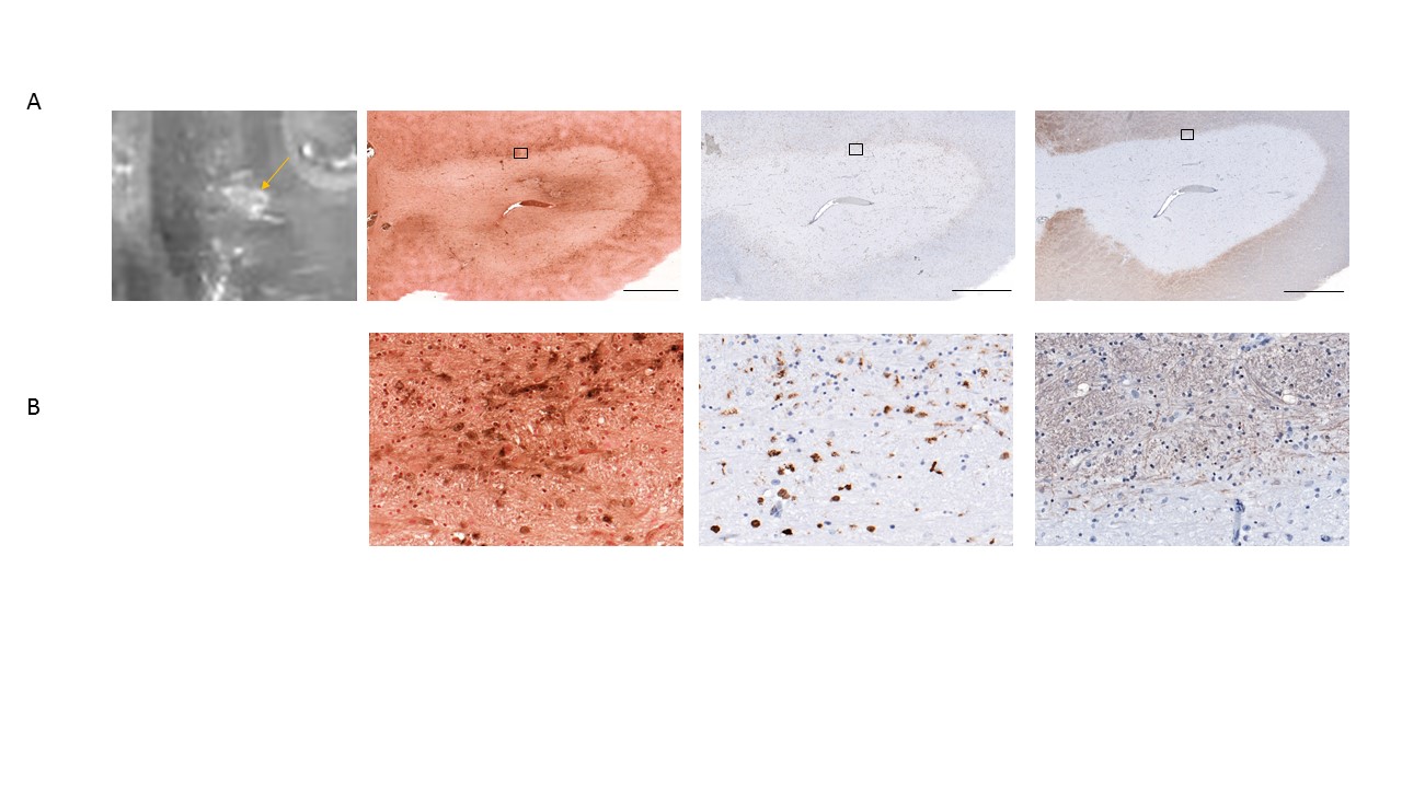
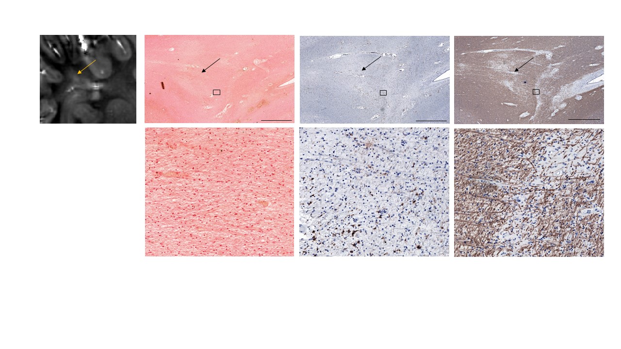
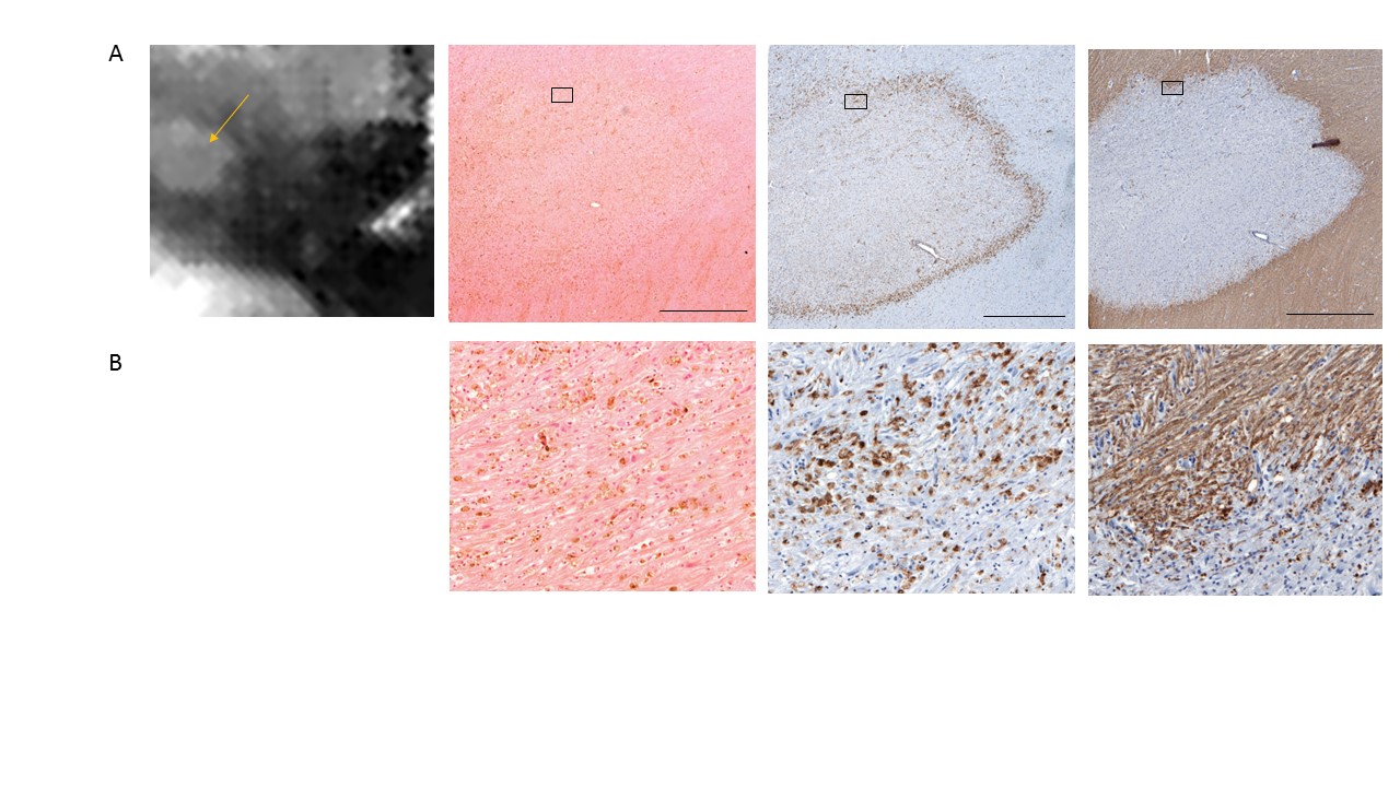
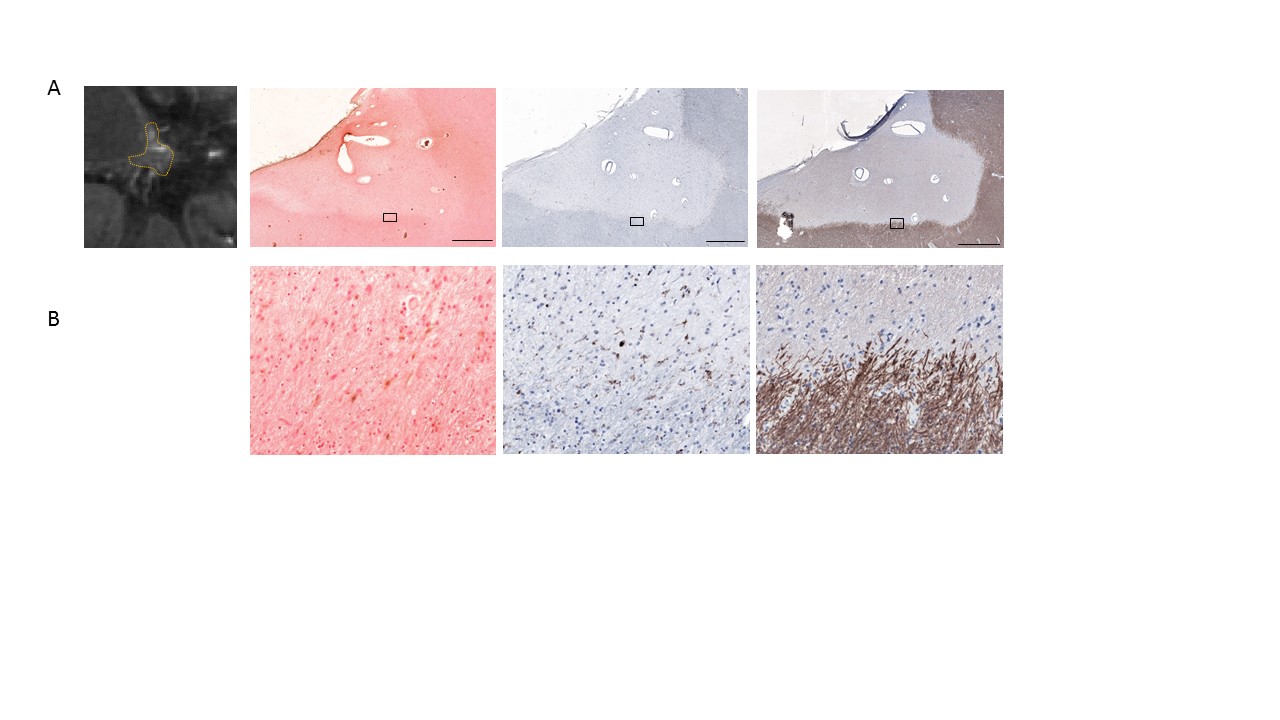
Brain slabs from patients with MS were obtained from the Rocky Mountain MS Center (RMMSC, Colorado, USA) and scanned on a 3T clinical MRI scanner (GE Healthcare, Wisconsin, USA) using an 8-channel coil. A T2FLAIR sequence (0.8 x 0.8 x 0.6 mm3, TE = 160 ms, TR = 9.6 s, bandwidth = ± 62.5 kHz) and 3D-gradient echo (GRE) sequence (0.6 x 0.6 x 0.6 mm3, TE1 = 4.3 ms, ΔTE = 4.8 ms, # TE = 11, TR = 74.2 ms, bandwidth = ± 62.5 kHz, 3 orientations) were used for QSM multiple orientation reconstruction (COSMOS) (3). After scanning, lesions were paraffin-embedded, sectioned into 5µm thick sections, and subjected to DAB-enhanced Perls’ stain (iron) and antibody labeling against myelin basic protein (MBP; myelin), and CD68 (macrophages and microglia). Slides were scanned with a digital scanner (Mirax).Results
Based on QSM of 28 lesions from post-mortem MS brain slabs, 6 were rim-positive (21%), 6 showed homogeneous signal throughout the lesion (21%), and 16 (58%) were non-homogeneous, as determined by a neuroradiologist. The frequency of rim-positive lesions is in agreement with recent results from MS patients in which 110 out of 514, or 21% of lesions were rim-positive (unpublished data).
Of the 6 rim-positive lesions, all were pro-inflammatory because they were chronic active lesions with iron-laden macrophages/microglia (Figure 1), and are therefore likely to be M1 polarized (2). The 6 homogeneous lesions were non-inflammatory: 5 lesions were chronic active and iron negative with varied loss of myelin (Figure 2), and 1 was an actively demyelinating lesion (Figure 3). The non-homogeneous lesions were also non-inflammatory, as they were predominantly chronic active and iron negative (Figure 4), chronic silent, or had iron positive scar tissue. Without excess iron, the macrophages/microglia present at the lesion rim are neither M1 nor M2 polarized (unpublished data), and are therefore not clinically concerning.
Discussion
We identified three susceptibility patterns on QSM where rim-positive lesions, but not other lesion types, were associated with iron-positive macrophages/microglia. Despite being chronically active, iron negative lesions are not clinically relevant because they have fewer macrophages/microglia, are not M1 polarized, and therefore do not carry out smoldering inflammation. Despite the absence of iron-positive macrophage/microglia, lesions still have hyperintense susceptibility. This signal is likely due to the presence of deoxygenated blood in vessels, astrocytic iron, and/or the incomplete loss of myelin.Conclusion
QSM identifies a subtype of MS lesion that contains iron and macrophages/microglia behind a sealed BBB. These lesions show a rim pattern and are clinically the most relevant as they are indicative of inflammatory activity and thus provide a biomarker for chronic inflammation in established lesions.Acknowledgements
No acknowledgement found.References
1. Eskreis-Winkler S, Zhang Y, Zhang J, et al. The clinical utility of QSM: disease diagnosis, medical management, and surgical planning. NMR Biomed 2017;30(4).
2. Mehta V, Pei W, Yang G, et al. Iron is a sensitive biomarker for inflammation in multiple sclerosis lesions. PLoS One 2013;8(3):e57573.
3. Liu T, Spincemaille P, de Rochefort L, Kressler B, Wang Y. Calculation of susceptibility through multiple orientation sampling (COSMOS): a method for conditioning the inverse problem from measured magnetic field map to susceptibility source image in MRI. Magn Reson Med 2009;61(1):196-204.
Figures